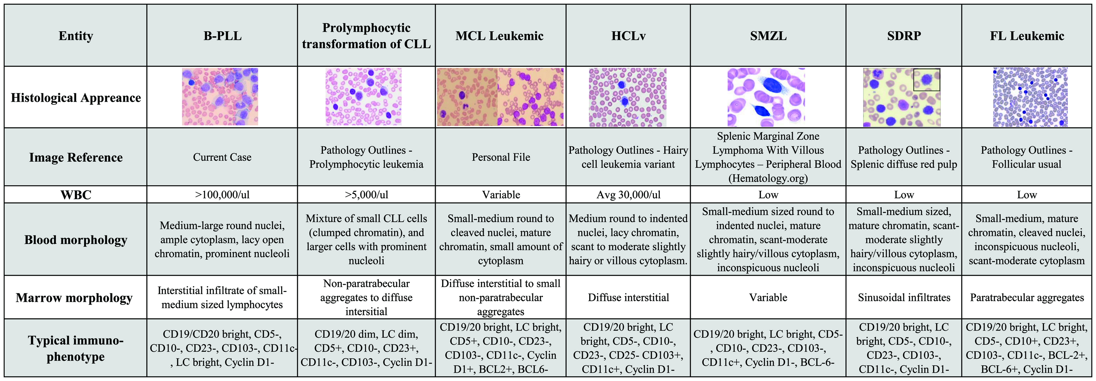
Figure 1. A broad differential diagnosis for B-PLL. B-PLL: B-cell prolymphocytic leukemia; CLL: chronic lymphocytic leukemia; MCL: mantle cell lymphoma; HCL-v: hairy cell leukemia variant; SMZL: splenic marginal zone lymphoma; SDRP: splenic diffuse red pulp; FL: follicular lymphoma; WBC: white blood cell; Avg: average; LC: light chain.
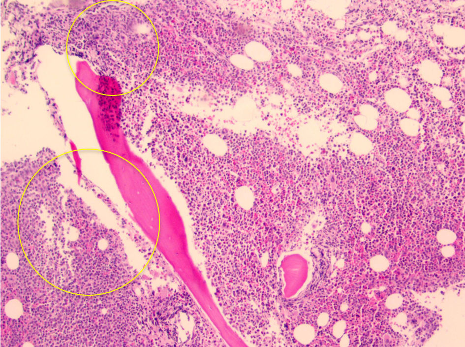
Figure 2. Core biopsy showing hypercellular marrow for age of patient (about 90%), with ill-defined aggregates of lymphocytes (yellow circled areas). Note the lack of “fried-egg” appearance which is seen with hairy cell leukemia.
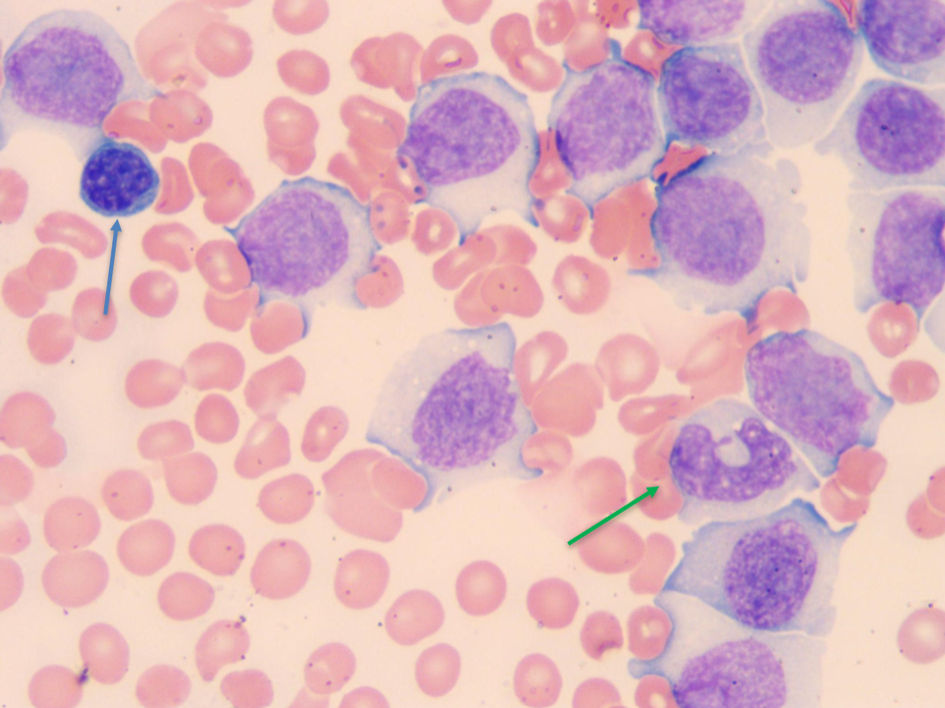
Figure 4. Blood smear showing a normal small lymphocyte near the upper left-hand corner (blue arrow), and normal monocytes near the lower right corner (green arrow) for comparison. The rest of the cells are B-PLL cells which are as big if not bigger than monocytes. B-PLL: B-cell prolymphocytic leukemia.