Journal of Hematology, ISSN 1927-1212 print, 1927-1220 online, Open Access
Article copyright, the authors; Journal compilation copyright, J Hematol and Elmer Press Inc
Journal website https://www.thejh.org

Case Report

Volume 12, Number 5, October 2023, pages 231-235


Cyclic Thrombocytopenia in the Setting of Intracranial Hemorrhage: A Diagnostic and Therapeutic Challenge

Figures

Figure 1.
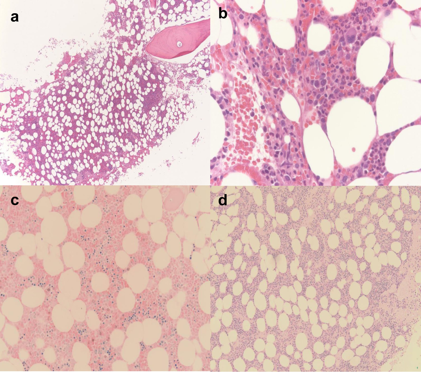
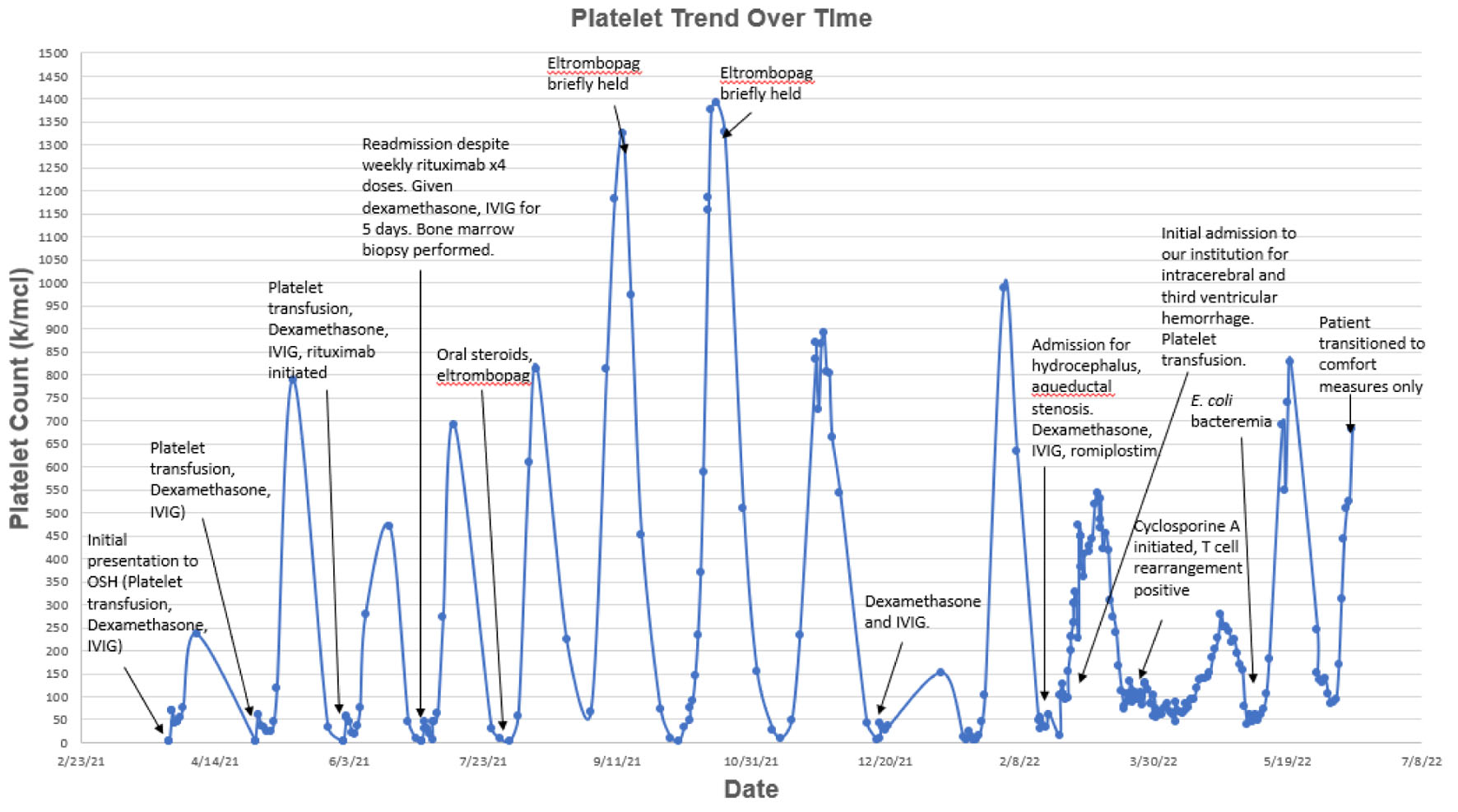
Figure 1. Platelet count over time. Patient presented to our institution with intracerebral and third ventricular hemorrhage at which time hematology was consulted. Looking back on prior platelet trends, patient had been hospitalized multiple times for recurrent episodes of severe thrombocytopenia. Upon pattern recognition, a diagnosis of cyclic thrombocytopenia was made and patient was initiated on cyclosporine A. Platelets stabilized to > 5.0 × 10⁴/µL, a threshold recommended by neurosurgery in the setting of ventriculoperitoneal shunt placement, for nearly 3 months.
Figure 2.
Figure 2. Bone marrow biopsy findings. (a) Low power × 2 magnification: the cellularity is 30%. Megakaryocytes are slightly decreased with minimal atypia. The myeloid to erythroid ratio appears unremarkable. (b) High power × 20: single megakaryocyte is visible with minimal atypia. (c) Giemsa stain: it shows normal myeloid to erythroid ratio. (d) Periodic acid-Schiff (PAS) stain: it shows normal myeloid to erythroid ratio.