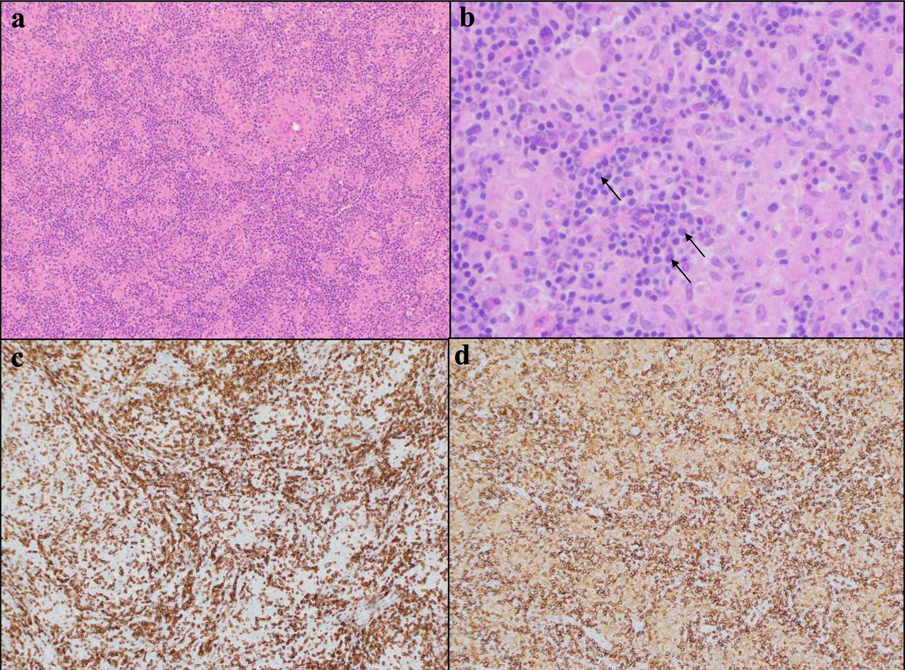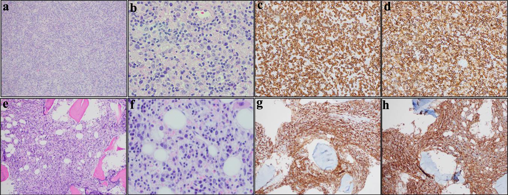
Figure 1. Cervical lymph node biopsy and bone marrow biopsy histology and immunohistochemistry confirming diagnosis of peripheral T-cell lymphoma, not otherwise specified (PTCL, NOS). (a)-(d) are cervical lymph node biopsy and (e)-(h) are bone marrow biopsy. (a) H&E of diagnostic lymph node at × 100. Clusters of epithelioid histiocytes have now been infiltrated by neoplastic lymphoid cells. (b) H&E of diagnostic lymph node at × 400. The arrows point to examples of large neoplastic T cells. (c) CD3 immunostain at × 100. (d) CD4 immunostain at × 400. The neoplastic T cells are CD4-positive/CD8-negative. (e) H&E of bone marrow at × 100. (f) H&E of bone marrow at × 400. (g) CD3 immunostain at × 100. (h) CD4 immunostain at × 400. H&E: hematoxylin and eosin.