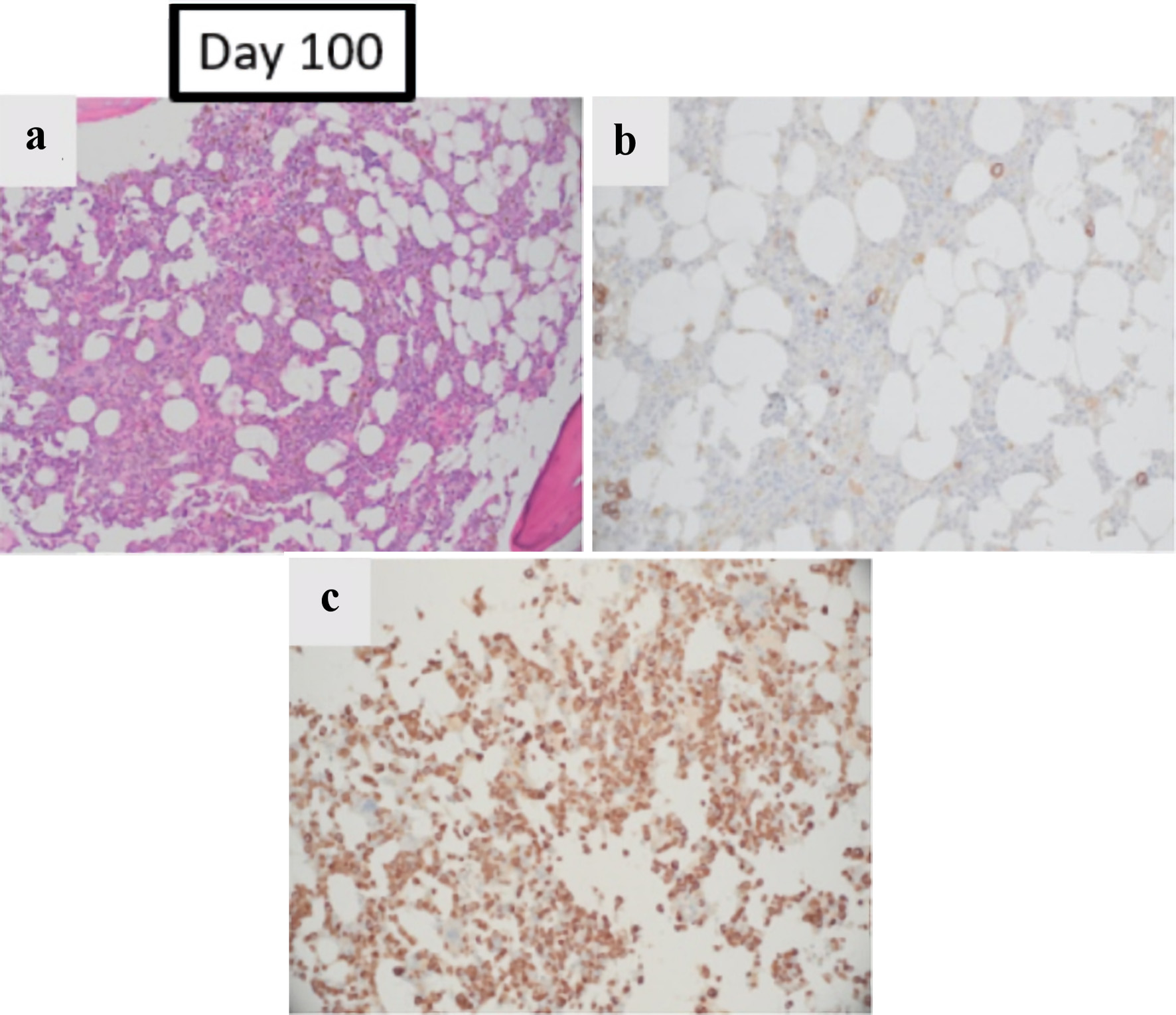
Figure 1. The effect of pure red cell aplasia on patient’s BM: BM obtained on day +100 post allo-HSCT, and prior to starting treatment for PRCA. (a) Hematoxylin and eosin stain (magnification: × 100) showing decreased erythroid elements. (b) E-cadherin stain which highlighted decreased erythroid precursors (< 5% of total cells) with limited colony formation (magnification: × 200). (c) Myeloperoxidase stain which showed marked predominance of maturing granulocytic precursors (magnification: × 200). Allo-HSCT: allogenic hematopoietic stem cell transplant; BM: bone marrow; PRCA: pure red cell aplasia.

