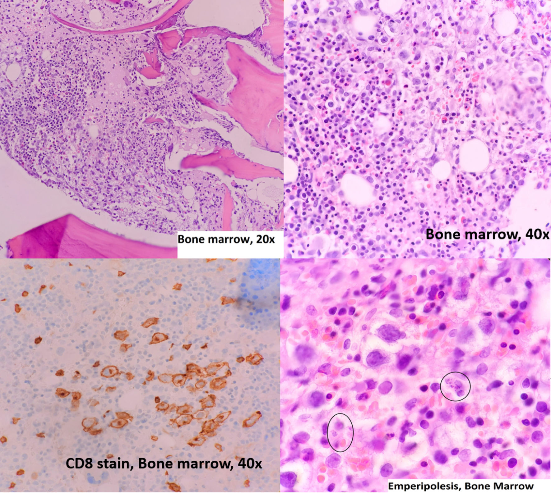
Figure 1. Bone marrow aspiration, hematoxylin and eosin stain and immunohistochemistry. Circles mark the emperipolesis process.
| Journal of Hematology, ISSN 1927-1212 print, 1927-1220 online, Open Access |
| Article copyright, the authors; Journal compilation copyright, J Hematol and Elmer Press Inc |
| Journal website https://www.thejh.org |
Case Report
Volume 13, Number 1-2, April 2024, pages 29-33
Hepatosplenic Alpha-Beta T-Cell Lymphoma: A Challenging Diagnostic Entity
Figures