Journal of Hematology, ISSN 1927-1212 print, 1927-1220 online, Open Access
Article copyright, the authors; Journal compilation copyright, J Hematol and Elmer Press Inc
Journal website https://www.thejh.org

Case Report

Volume 13, Number 3, June 2024, pages 94-98


Non-Secretory Multiple Myeloma Associated With High-Risk Phenotype and Complex Cytogenetics Including t(8;22)

Figures

Figure 1.
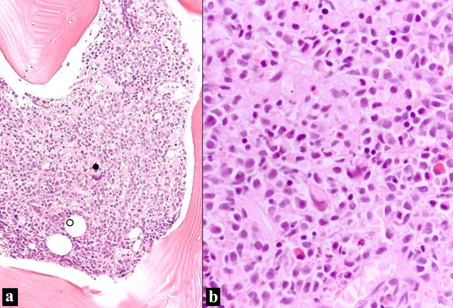
Figure 1. Bone marrow biopsy (H&E stain). (a) Bone marrow intertrabecular space diffusely infiltrated by sheets of non-cohesive cells. Residual bone marrow elements are scattered (• megakaryocyte; ○ adipocytes) (H&E, × 100 optical magnification). (b) Sheet of atypical plasma cells infiltrating with residual bone marrow elements. The plasma cells have eccentric nuclei with mild atypia and moderate eosinophilic cytoplasm with perinuclear hofs (H&E, × 400). H&E: hematoxylin and eosin.
Figure 2.
Figure 2. Bone marrow biopsy (IHC and ISH). (a) Plasma cells show strong membranous staining (CD138, IHC, × 100). (b) Few scattered positive cells (blue) represent residual uninvolved plasma cells; the atypical plasma cells appear negative (kappa light chain, ISH, × 100). (c) Few scattered positive cells (blue) represent residual uninvolved plasma cells; the atypical plasma cells appear negative (lambda light chain, ISH, × 100). IHC: immunohistochemical; ISH: in situ hybridization.
Figure 3.
Figure 3. MYC break apart probe: t(8;22)(q24.2;q11.2) detected using dual color dual fusion FISH probe. FISH: fluorescence in situ hybridization.