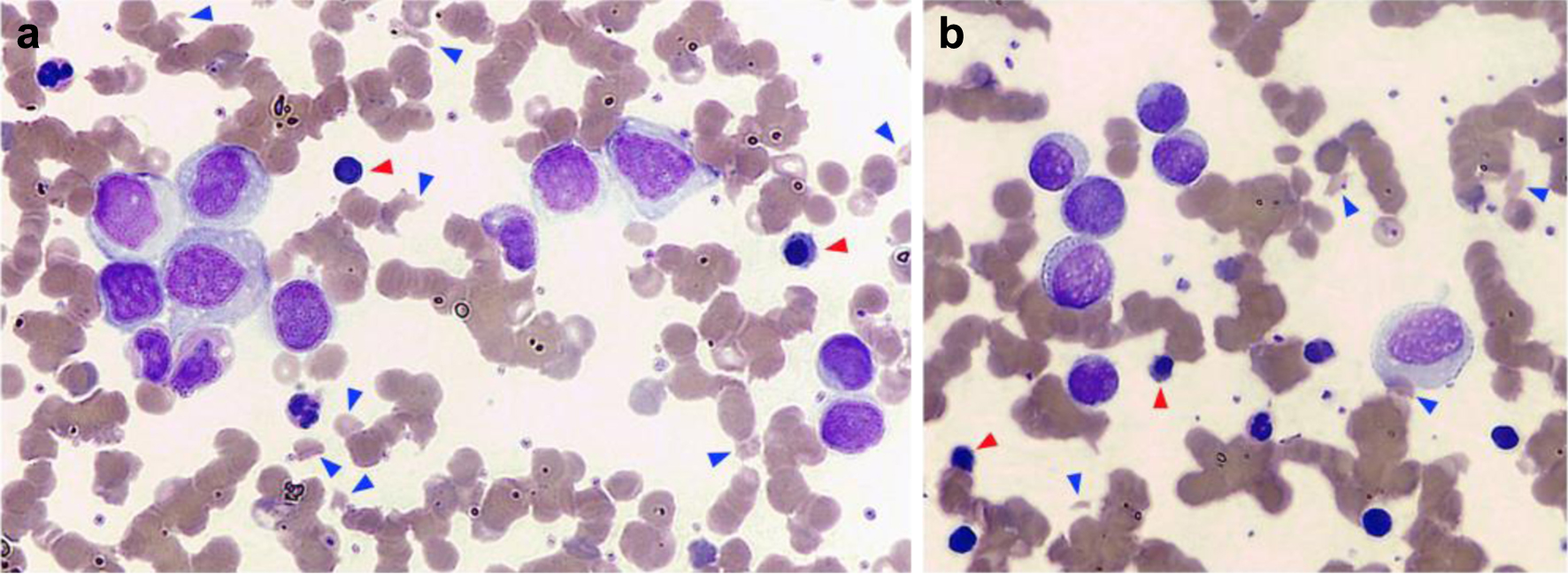
Figure 1. Peripheral blood smear. (a) Marked red cell anisocytosis with a remarkable proportion of schistocytes (15% of erythrocytes, blue arrowheads) and erythroblasts (3% of nucleated cells, red arrowheads) was observed. (b) A significant number of large atypical mononuclear cells with eccentric nucleus, of uncertain lineage, were also noted (15% of nucleated cells).
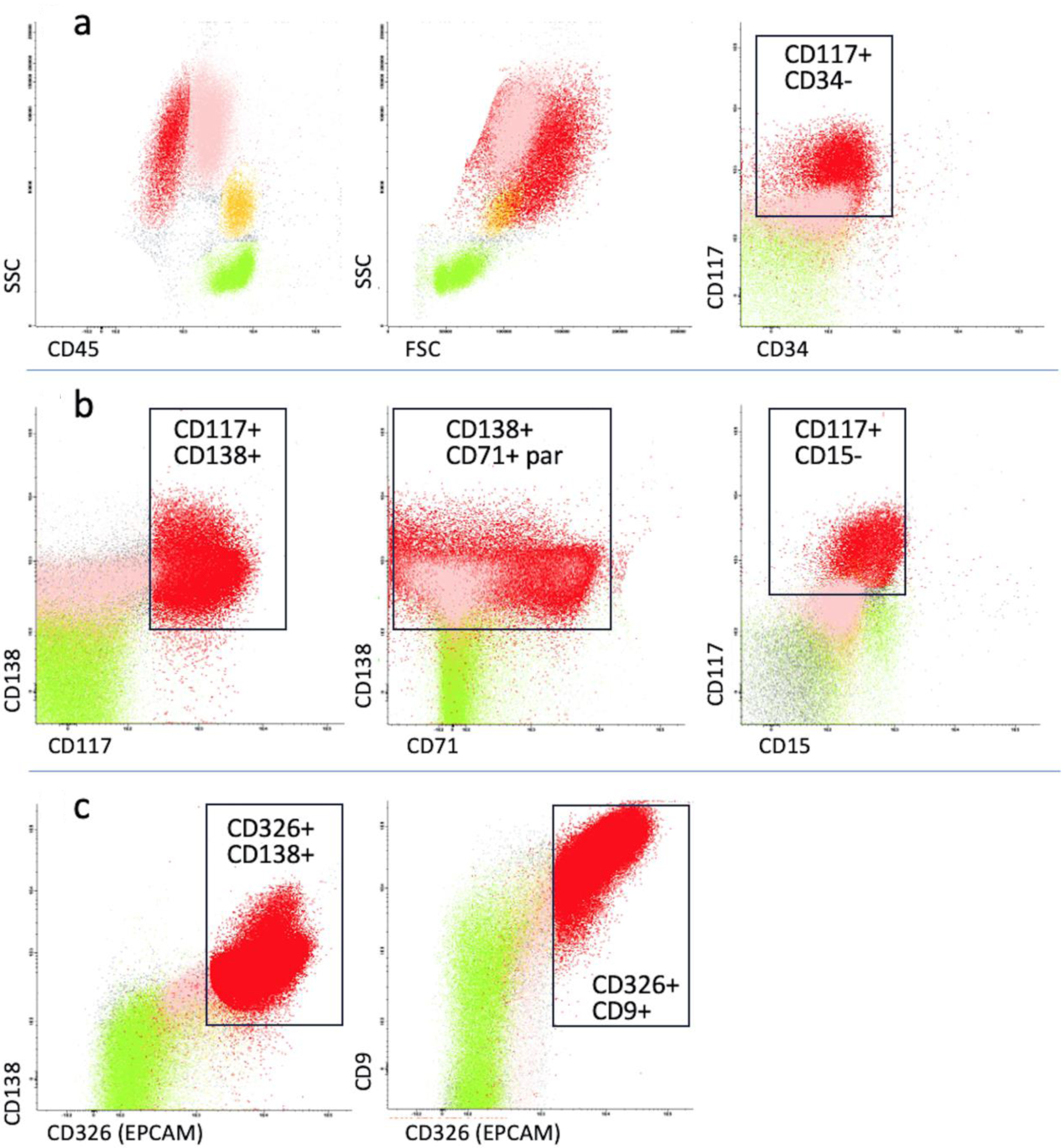
Figure 2. Immunophenotypic analysis of peripheral blood. Green: lymphocytes; yellow: monocytes; pink: neutrophils; red: circulating breast cancer cells. (a) The first myeloid flow cytometry panel showed an atypical population (red) of 20%, negative for CD45 and CD34, positive for CD117 with high forward and side scatter. (b) A more extensive panel included plasma cell (CD138, CD38, CD19) and erythroid markers (CD71). The atypical population resulted positive for CD138 and CD71 with negativity for CD38. (c) A panel for non-hematological markers including CD326 epithelial cell adhesion molecule (EpCAM) was carried out. The abnormal population expressed EpCAM and CD9. The black rectangles indicate the areas of positive expression of cell markers, where applicable. FSC: forward scatter; SSC: side scatter; par: partial expression.
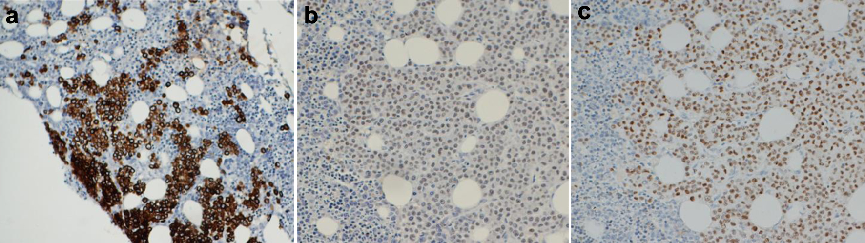
Figure 3. Immunohistochemistry analysis of bone marrow biopsy. A massive infiltration of cancer cells expressing all epithelial markers identified at diagnosis, such as cytokeratin (a), GATA-binding protein 3 (GATA3) (b) and estrogen receptor (c) was demonstrated.