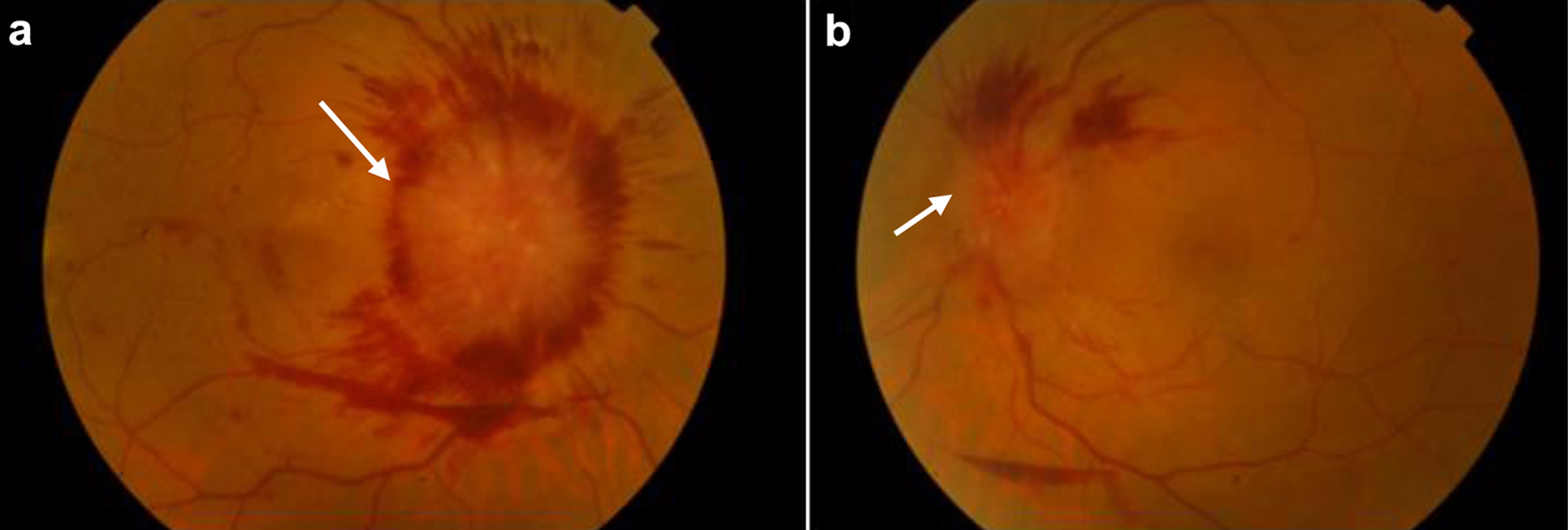
Figure 1. (a) Color fundus photograph of the right eye, showing extensive optic disc swelling (white arrow) with flame-shaped retinal hemorrhages around the optic disc, as well as dot-blot hemorrhages at the foveal area and a pre-retinal hemorrhage inferior of the optic disc. (b) Color fundus photography of the left eye, showing optic disc swelling (white arrow) and flame-shaped retinal hemorrhages superior to the optic disc, as well as a small pre-retinal hemorrhage adjacent to the inferior arcade.
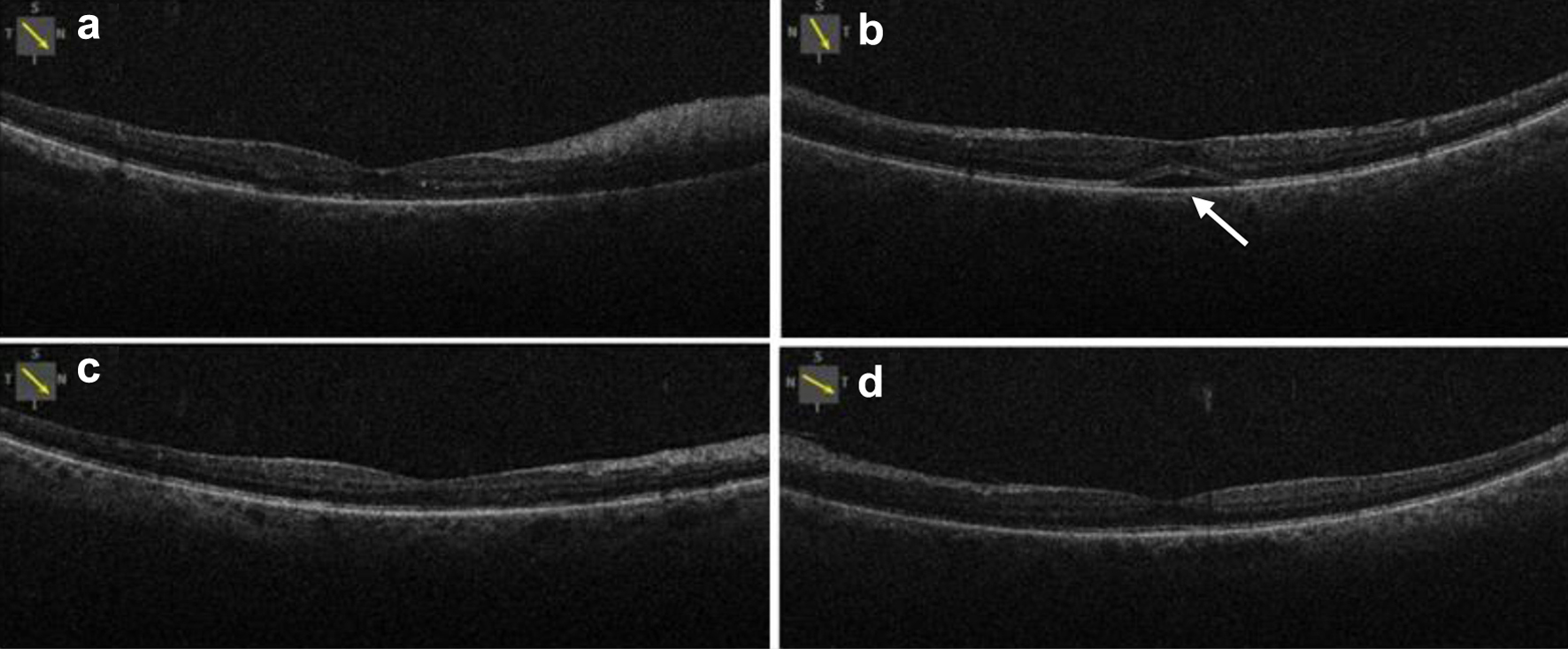
Figure 2. Optical coherence tomography of the right (a) and left (b) eye, showing normal macula in the right eye and small serous retinal detachment (white arrow) in the left eye at baseline. Optical coherence tomography of the right eye (c) and left (d) eye, showing normal retinal in both eyes with absorption of fluid in the left eye.
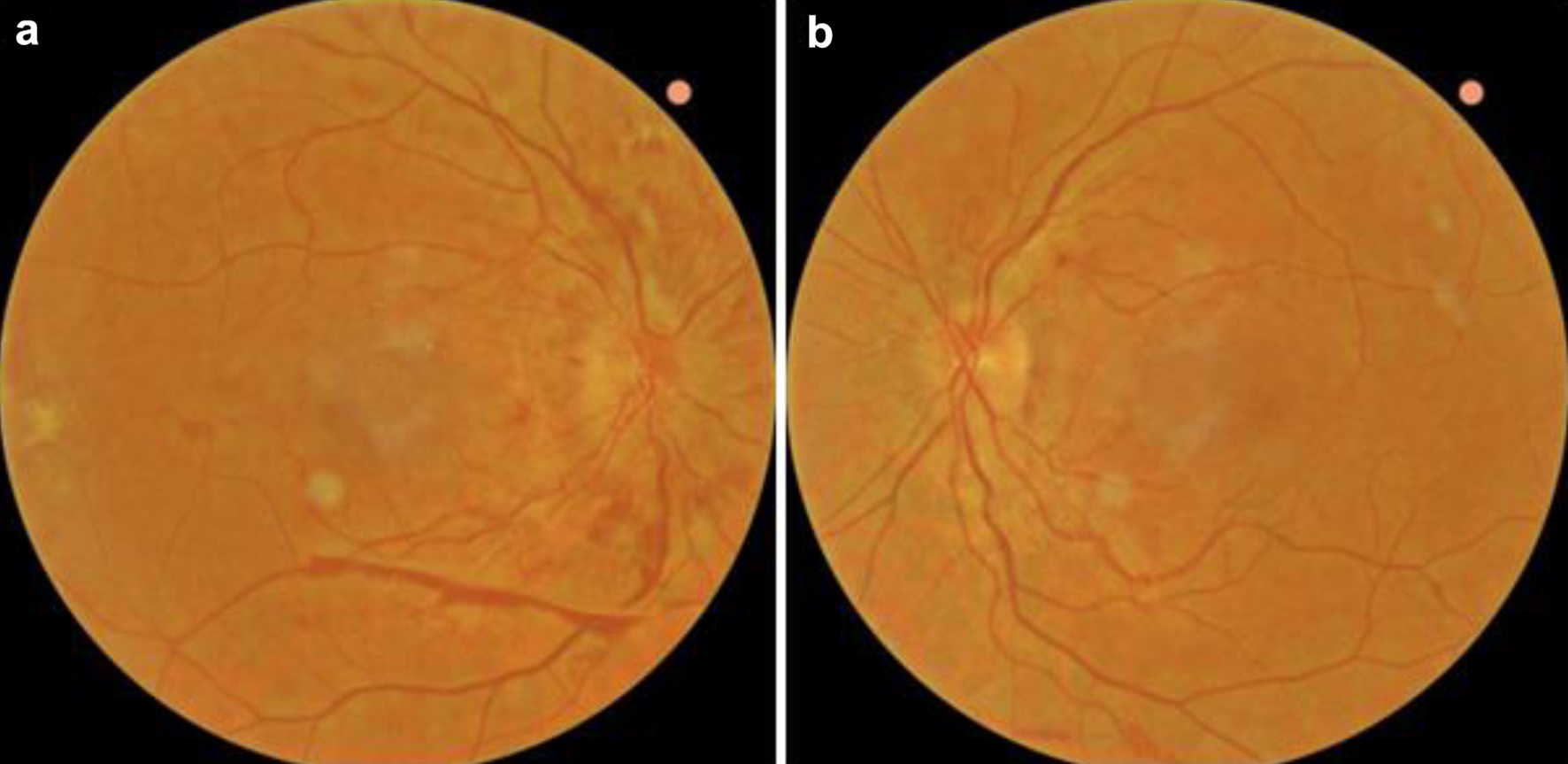
Figure 3. Color fundus photographs of the right (a) and left (b) eye 2 months after initiation of chemotherapy, showing improvement of the optic disc swelling in both eyes and almost full regression of retinal hemorrhages.