Journal of Hematology, ISSN 1927-1212 print, 1927-1220 online, Open Access
Article copyright, the authors; Journal compilation copyright, J Hematol and Elmer Press Inc
Journal website https://www.thejh.org

Case Report

Volume 13, Number 3, June 2024, pages 128-136


Unraveling the Rare Entity of KIT D816V-Negative Systemic Mastocytosis

Figure

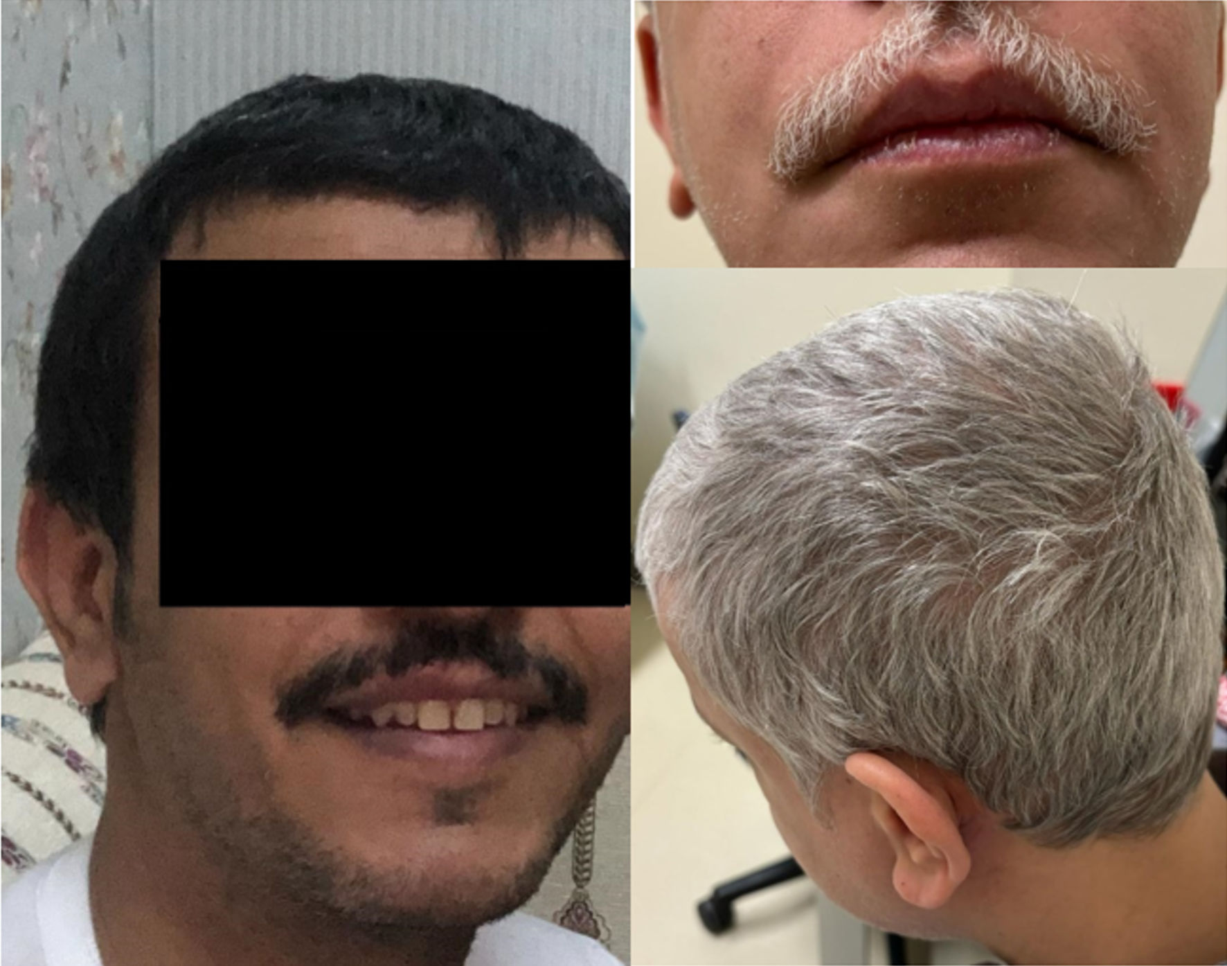
Figure 1.
Figure 1. Case 2: skin and hair color pre (left) and post (right) avapritinib treatment. Wild-type KIT signaling is involved in melanogenesis and hair pigmentation, and inhibition can result in hair depigmentation and lightning of the skin through a temporary melanocyte dysfunction. Avapritinib skin and hair changes were reported in 6% to 21% of treated patients.

Tables

Table 1. The WHO Fifth Edition (WHO 5th) and the ICC Classifications of Mastocytosis
 
Cutaneous mastocytosisICCWHO 5th
aIn the WHO classification, well-differentiated systemic mastocytosis (WDSM) is a morphologic variant that can occur in any SM subtype, including mast cell leukemia. bIn the ICC, those variants of SM must be correlated with B- and C-findings. cIn the ICC, mast cell leukemia must meet the diagnostic criteria of SM and have ≥ 20% atypical immature mast cells. dBone marrow mastocytosis (BMM) typically lacks the B-findings and with normal/near normal serum tryptase. The WHO classifies BMM as an SM subcategory, while in ICC, it is considered a variant of indolent SM. SM: systemic mastocytosis; WHO: World Health Organization; ICC: International Consensus Classification.
1. Urticaria pigmentosa/maculopapular cutaneous mastocytosis1. Urticaria
2. Diffuse cutaneous mastocytosis  Pigmentosa/maculopapular cutaneous mastocytosis
3. Mastocytoma of skin    Monomorphic
    Polymorphic
2. Diffuse cutaneous mastocytosis
3. Cutaneous mastocytoma
  Isolated mastocytoma
  Multilocal mastocytoma
SMICCWHO 5tha
1. Indolent SM (includes bone marrow mastocytosis)b1. Bone marrow mastocytosisd
2. Smoldering SMb2. Indolent SM
3. Aggressive SMb3. Smoldering SM
4. SM with an associated myeloid neoplasm4. Aggressive SM
5. Mast cell leukemiac5. SM with an associated hematologic neoplasm
6. Mast cell leukemia
Mast cell sarcomaICCWHO 5tha


Table 2. The WHO Fifth Edition (WHO 5th) and the ICC Diagnostic Criteria for SM
 
WHO 5thaICCa
aFor SM diagnosis at least one major and one minor or three minor criteria must be met. bSpindle-shaped mast cells do not count as an SM criterion when they line vascular cells, fat cells, nerve cells, or the endosteal-lining cell layer. Valent et al. describe detailed morphological criteria [11]. cFor KIT mutation, high-sensitivity PCR is recommended to avoid false negatives. In the absence of a KIT D816V mutation, exclusion of KIT mutation variants is strongly recommended (this includes codons 417, 501 - 509, 522, 557 - 560, 642, 654, 799, 816, 820, 822, the list of variants provided in the appendix of WHO proposal [1]. In cases of negative KIT, particularly those with eosinophilia, tyrosine kinase gene fusions associated with myeloid/lymphoid neoplasms with eosinophilia and tyrosine kinase gene fusions must be excluded. dBy flow cytometry or immunohistochemistry or by both techniques. eIn the WHO classification, patients with hereditary alpha-tryptasemia (HαT) need adjustment for tryptase level (although not standardized). The suggested correction is to divide the basal tryptase level by (1 + the extra copy numbers) of the alpha tryptase gene. For example, when the tryptase level is 60 and three extra copies of the alpha tryptase gene are present, the corrected tryptase level is 15 (60/4 = 15) and thus it does not meet this minor SM criterion. fPer ICC, elevated tryptase does not count as a minor SM criterion in SM with an associated myeloid neoplasm. SM: systemic mastocytosis; WHO: World Health Organization; ICC: International Consensus Classification; BM: bone marrow.
Major criterion
Infiltrates of mast cells (≥ 15 mast cells in aggregates) in BM or extracutaneous organsbInfiltrates of tryptase- and/or CD117 positive mast cells (≥ 15 mast cells in aggregates) in the BM or extracutaneous organsb
Minor criteria
≥ 25% of all mast cells are atypical cells (type I or type II) or are spindle-shaped≥ 25% of all mast cells are atypical cells or are spindle-shaped
KIT-activating KIT point mutation(s) at codon 816 or in other critical regions of KITcKIT-activating KIT point mutation(s) at codon 816 or in other critical regions of KITc
Mast cells express at least one of the following markers: CD2 and/or CD25 and/or CD30dMast cells express at least one of the following markers: CD2 and/or CD25 and/or CD30d
Baseline serum tryptase > 20 ng/mLeBaseline serum tryptase > 20 ng/mLf


Table 3. The WHO Fifth Edition Summarizing the Clinical Findings of SM, Classified Into B-Findings and C-Findings (Adapted From Valent et al [1, 9])
 
B-findingsC-finding
WHO: World Health Organization; BM: bone marrow; VAF: variant allele frequency; PB: peripheral blood; MPN: myeloproliferative neoplasm; MDS: myelodysplastic syndrome; LN: lymph node; ANC: absolute neutrophil count; Plt: platelet; Hb: hemoglobin; HTN: hypertension; GIT: gastrointestinal tract.
Increased mast cell burden: ≥ 30% by histology on BM and/or serum tryptase ≥ 200 ng/mL and/or KIT D816V VAF ≥ 10% in BM or PB white blood cellsCytopenia/s (one or more of the following): ANC < 1 × 10⁹/L, Hb < 10 g/dL, Plt < 100 × 109/L
Features suggestive of MPN and/or MDSHepatopathy: hepatomegaly or cirrhosis ± ascites and elevated liver enzymes ± portal HTN
Organomegaly: palpable hepatomegaly or/and splenomegaly without systemic effects (i.e., no ascites or signs of organ damage and no hypersplenism, weight loss, and/or lymphadenopathy or visceral LN enlargement (> 2 cm) on imagingSpleen: palpable splenomegaly with hypersplenism ± weight loss ± hypoalbuminemia
GIT: malabsorption and hypoalbuminemia ± weight loss
Bone: large-sized osteolysis (≥ 2 cm) with pathologic fracture ± bone pain


Table 4. Clinical and Pathologic Comparison of Our SM Cases and Cases in the Literature
 
SexAge (years)SM subtypePresenting symptomsManagementResponse
aThe patient died of hemorrhagic stroke and disseminated intravascular coagulation. bThe patient initially responded to interferon-α, however, it was changed due to myelosuppression. Cladribine resulted in improvement in eosinophilia. The patient died from sepsis. SM: systemic mastocytosis.
Case 1F49Aggressive SMSkin rashes, dizziness, nausea, vomitingImatinib 300 mgClinical response
Case 2M40Indolent SMRecurrent anaphylactic episodesAvapritinib 150 mg/dayClinical response
Azad et al, 2023 [25]M53Aggressive SMHeadache, fatigue, weight loss, cognitive impairment, insomnia, exertional dyspnea, abdominal pain, bone and joint pain, diarrhea, bloody stools, and a pruritic rash localized in the torso and extremitiesFailed imatinib, cladribine, nilotinib, hydroxyurea, and midostaurinClinical and biochemical response to avapritinib.
Avapritinib 200 mg/day
Conde-Fernandes et al, 2017 [27]M15Indolent SMRecurrent flushing, hypotension, and syncope, preceded by skin lesionsOral disodium cromoglycateClinical and biochemical response
Caceres-Nazario et al, 2016 [14]M78Aggressive SMIncidental discovery of normocytic anemia with thrombocytopeniaImatinib to interferon-αDeath
Prednisone
Savini et al, 2015 [28]aF65Mast cell leukemiaFlushing and hypotensionAntihistamineDeath
Imatinib 400 mg to dasatinib 100 mg
Sakane-Ishikawa et al, 2013 [26]bF43Aggressive SMVertebral compression fracture, multiple lytic bone lesions with epidural mass, and eosinophilsEmergent laminectomy and subsequent irradiation of the tumorDeath
Interferon-α to dasatinib to cladribine