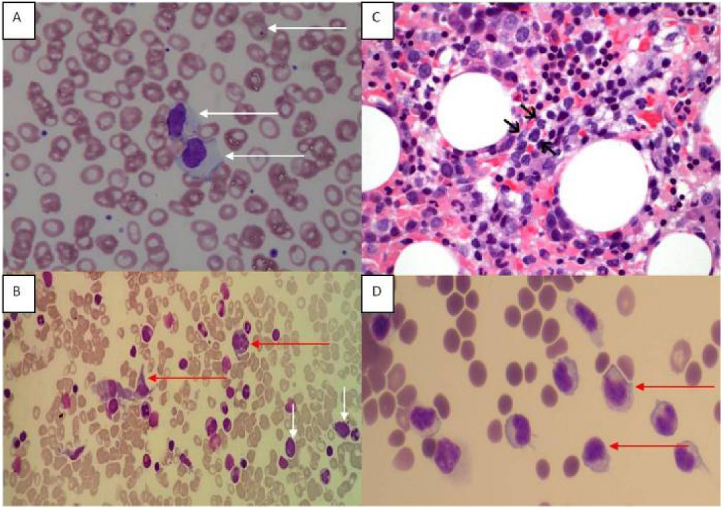
Figure 1. (A) Peripheral blood film showing large granular lymphoid cells. Howell Jolly bodies are seen in some red cells in keeping with a post-splenectomy picture. (B) Bone marrow aspirate (MGG stain) showing hemophagocytic activity and atypical lymphoid cells. Red arrows point to histiocytes, and white arrows indicate atypical lymphoid cells. (C) Bone marrow trephine showed a sinusoidal infiltrate of small to medium sized lymphocytes (arrows) with irregular nuclei and indistinct nucleoli (H&E, original magnification, × 600). (D) Ascitic fluid showing atypical lymphoid cells.
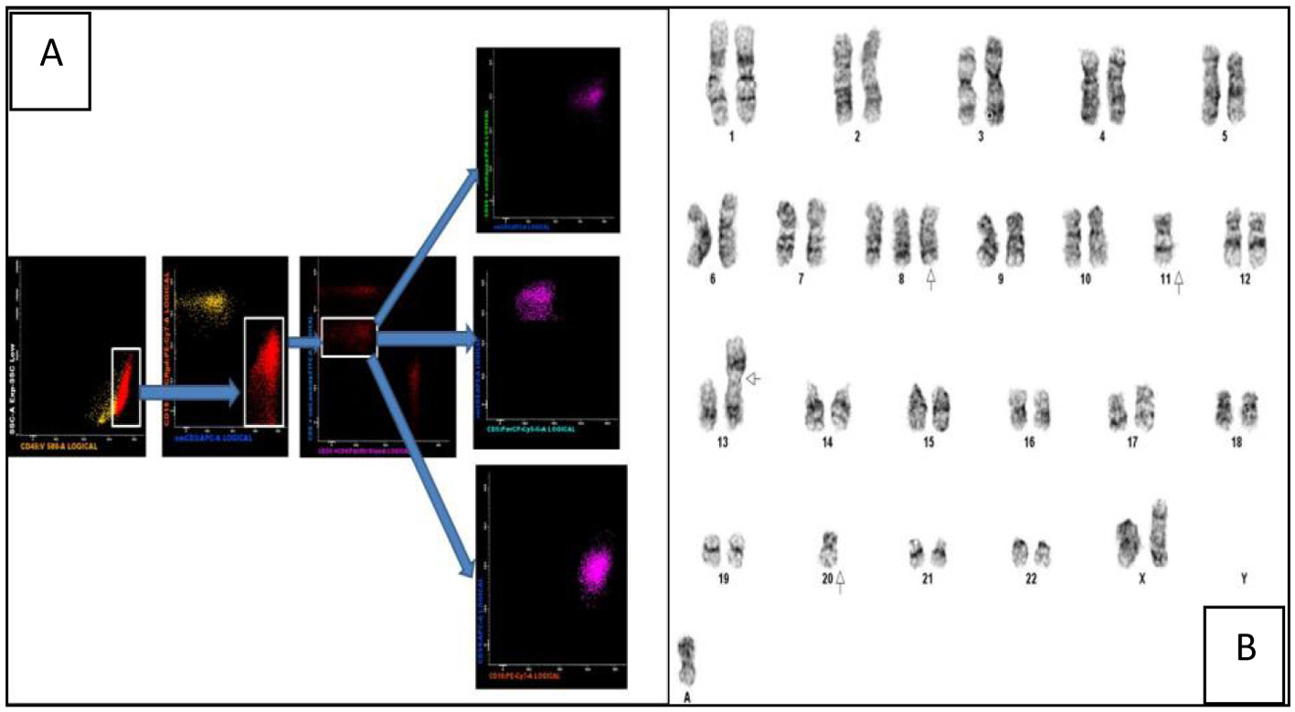
Figure 2. (A) Multiparametric flow cytometric analysis of the bone marrow specimen. The abnormal events are CD45+ with a low side scatter. They are T cells as evidenced by CD3 positivity and lack of CD19 expression (the yellow events in the CD19/CD3 scatter plot are normal B cells). Among the CD8+ T cells there is an abnormal CD8dim sub-population which constitutes the neoplastic clone. These events are CD5-/CD56+/CD16+ and CD94-. (B) Karyogram showing trisomy 8, loss of chromosomes 11 and 20, a non-clonal abnormality of chromosome 13 and a marker chromosome.
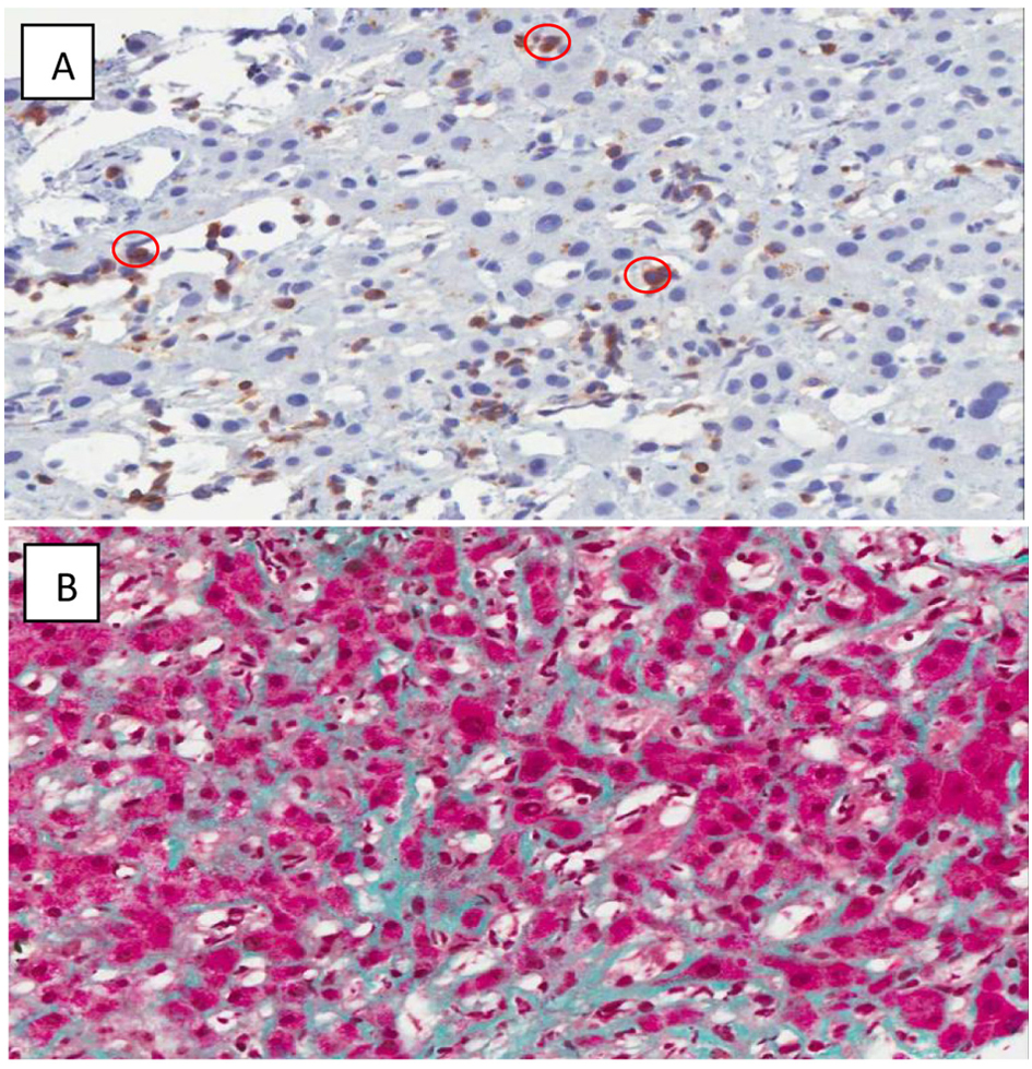
Figure 3. (A) CD 3 Immunoperoxidase highlights the T-cells circled in red. (× 400) Some T-cells are somewhat larger than others. (B) Sinusoidal fibrosis. (Masson's trichrome stain, × 200) The hepatocytes are red, and the collagen is green. Collagen is normally restricted to portal tracts and the cuff of central veins, but here is seen running along sinusoids between plates of hepatocytes