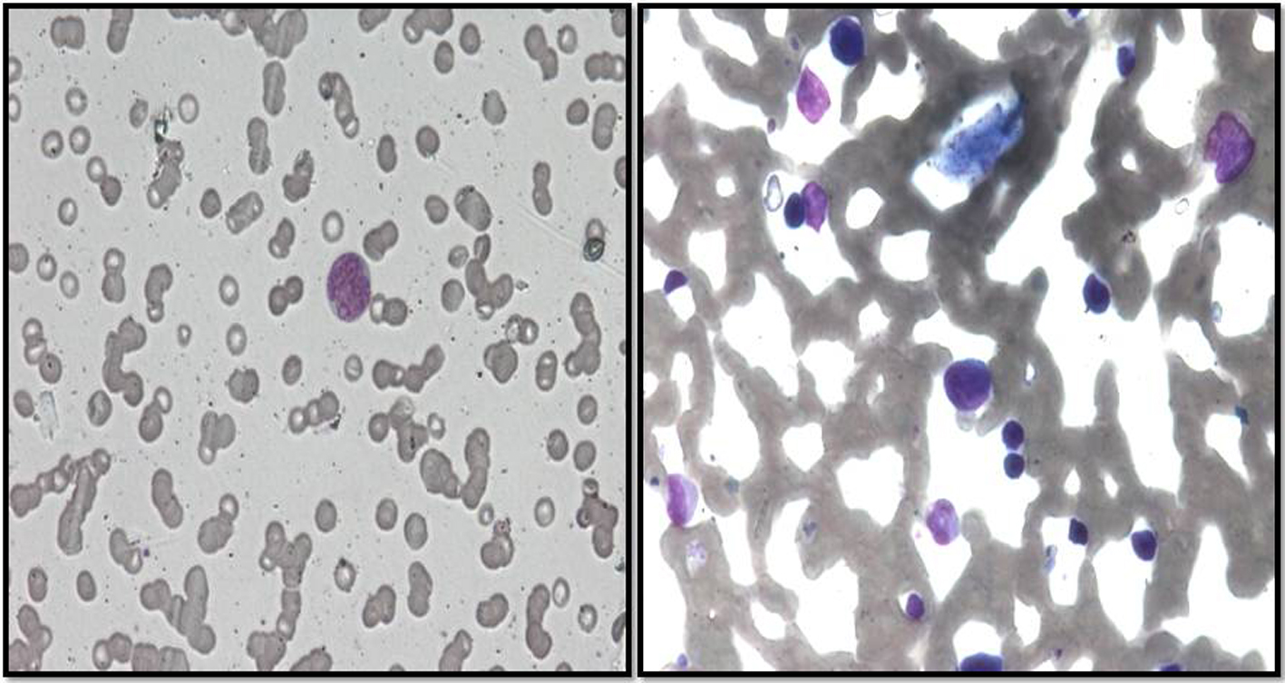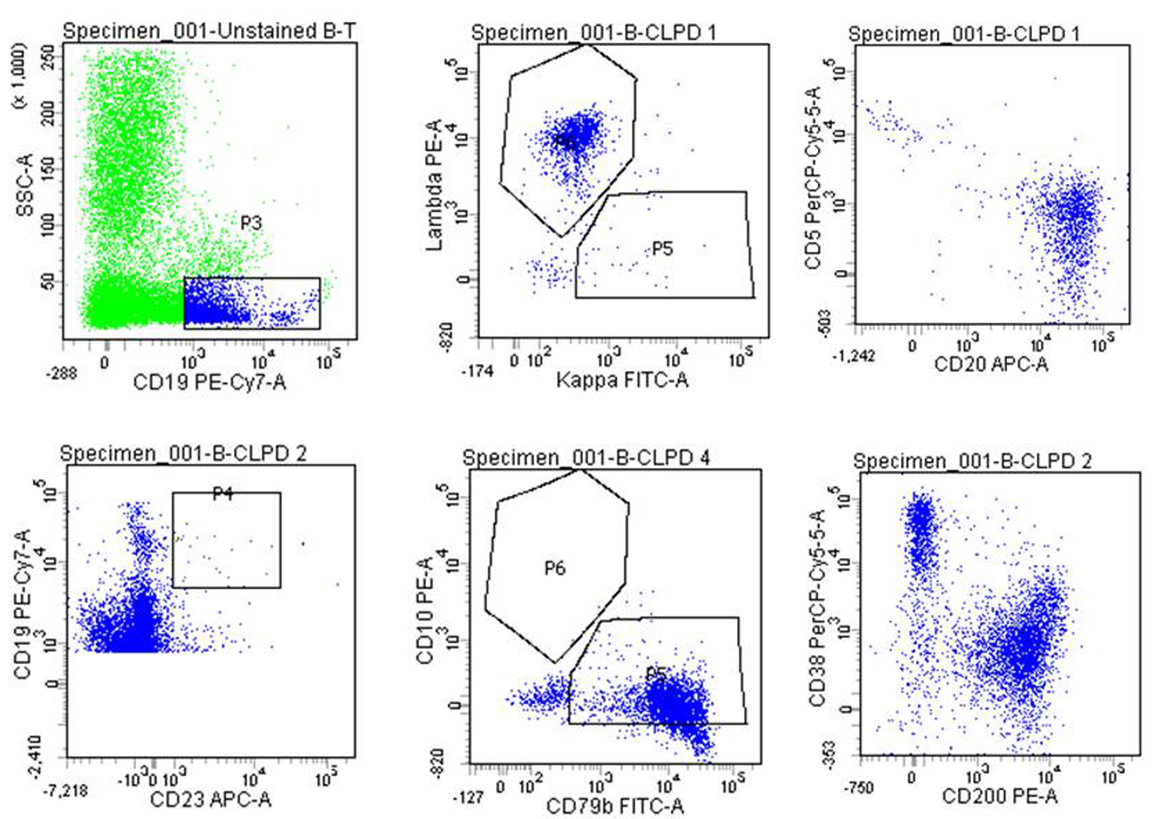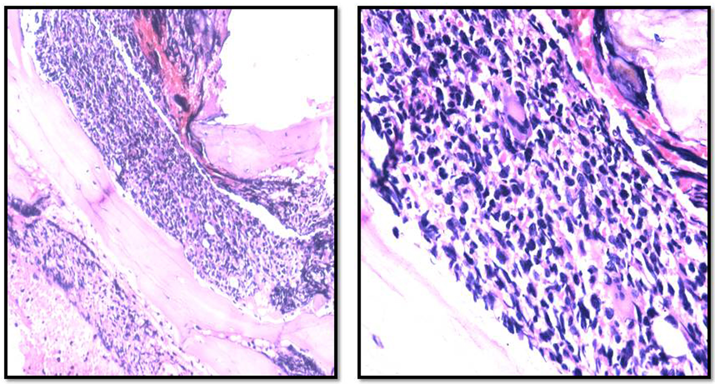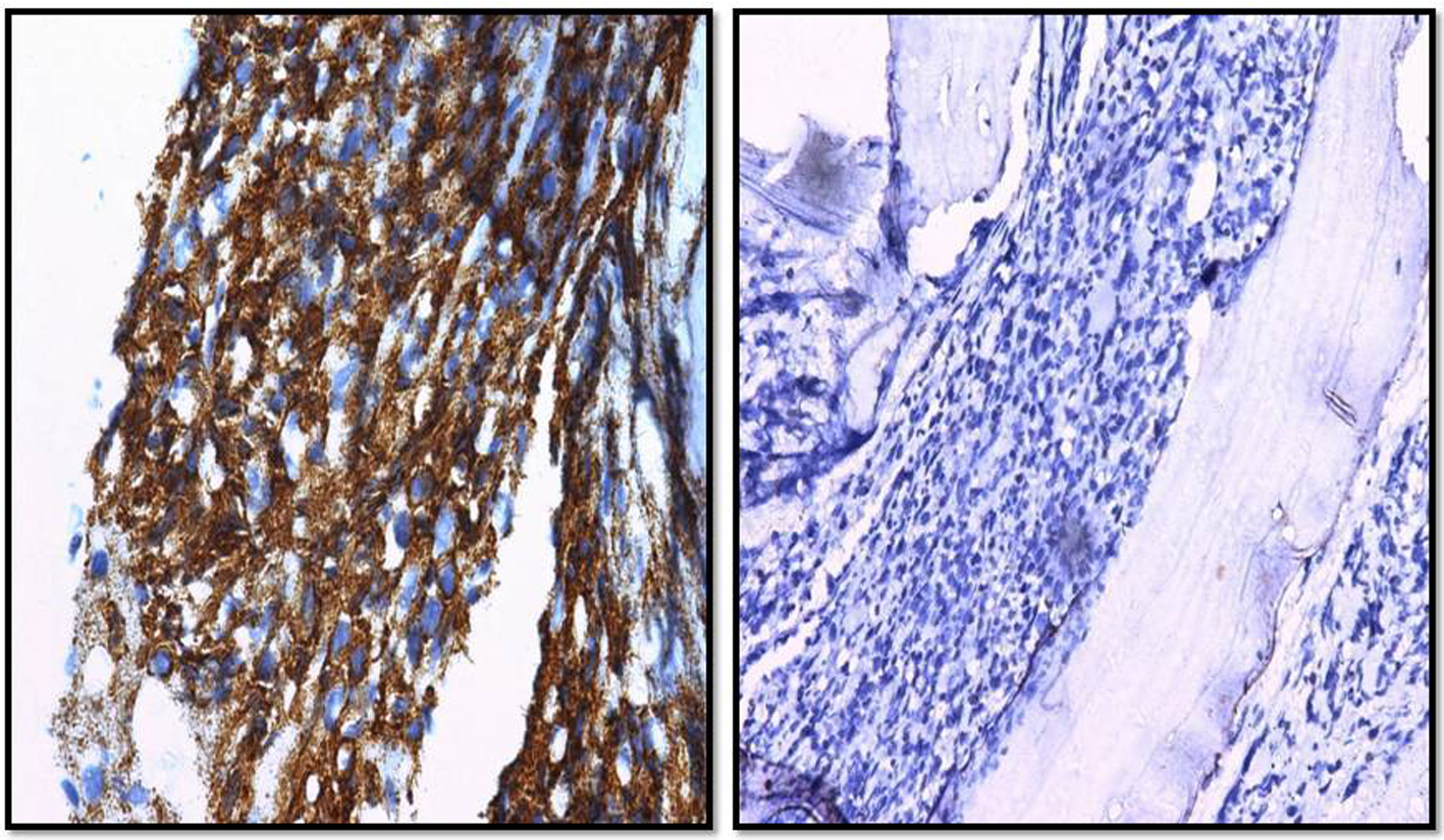
Figure 1. Peripheral blood and bone marrow aspirate revealed large sized atypical lymphoma cells with indistinct nucleoli and basophilic cytoplasm (Leishman stain, × 200).
| Journal of Hematology, ISSN 1927-1212 print, 1927-1220 online, Open Access |
| Article copyright, the authors; Journal compilation copyright, J Hematol and Elmer Press Inc |
| Journal website http://www.thejh.org |
Case Report
Volume 4, Number 4, December 2015, pages 242-245
Primary Bone Marrow B-Cell Lymphoma: Correlation of Results of Flow Cytometry and Morphological Findings
Figures


