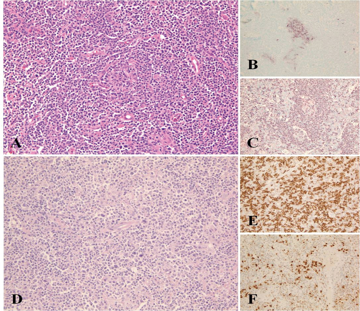
Figure 1. Morphological appearance of Castleman disease diagnosed at initial lymph node sampling. Compressed and partially hyalinized germinal centre surrounded by mantle zone B-cells HE, (A). Residual GC are highlighted by CD10 immunostaining of the same region (B) and remain negative by CD20 due to involution and hialinization (C). Diffuse large B-cell lymphoma showing solid areas of large CD20+ cells (D, E). MUM1 positivity supports the activated B-cell origin of the change (F).
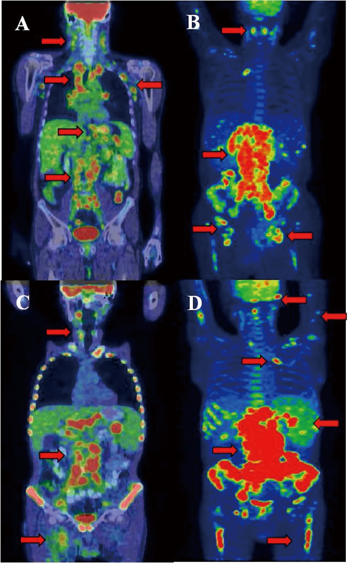
Figure 2. Castleman disease showed activity in many lymphatic areas (A). During staging of DLBCL significant progression was detected in the infradiagphragmatic region with bone marrow involvement (B). After 2 cycles of R-CHOP interim PET/CT proved improvement in the supradiaphragmatic, but not in the infradiaphragmatic region. After several chemotherapeutic protocols refractory state was confirmed by PET/CT and clinical signs (D). Arrows show the involved lymph nodes, spleen, lung and bone marrow. Arrows shows tracer enhanced areas.