Journal of Hematology, ISSN 1927-1212 print, 1927-1220 online, Open Access
Article copyright, the authors; Journal compilation copyright, J Hematol and Elmer Press Inc
Journal website http://www.thejh.org

Original Article

Volume 7, Number 2, May 2018, pages 43-50


Retrospective Study of High Hemoglobin Levels in 56 Young Adults

Figures

Figure 1.
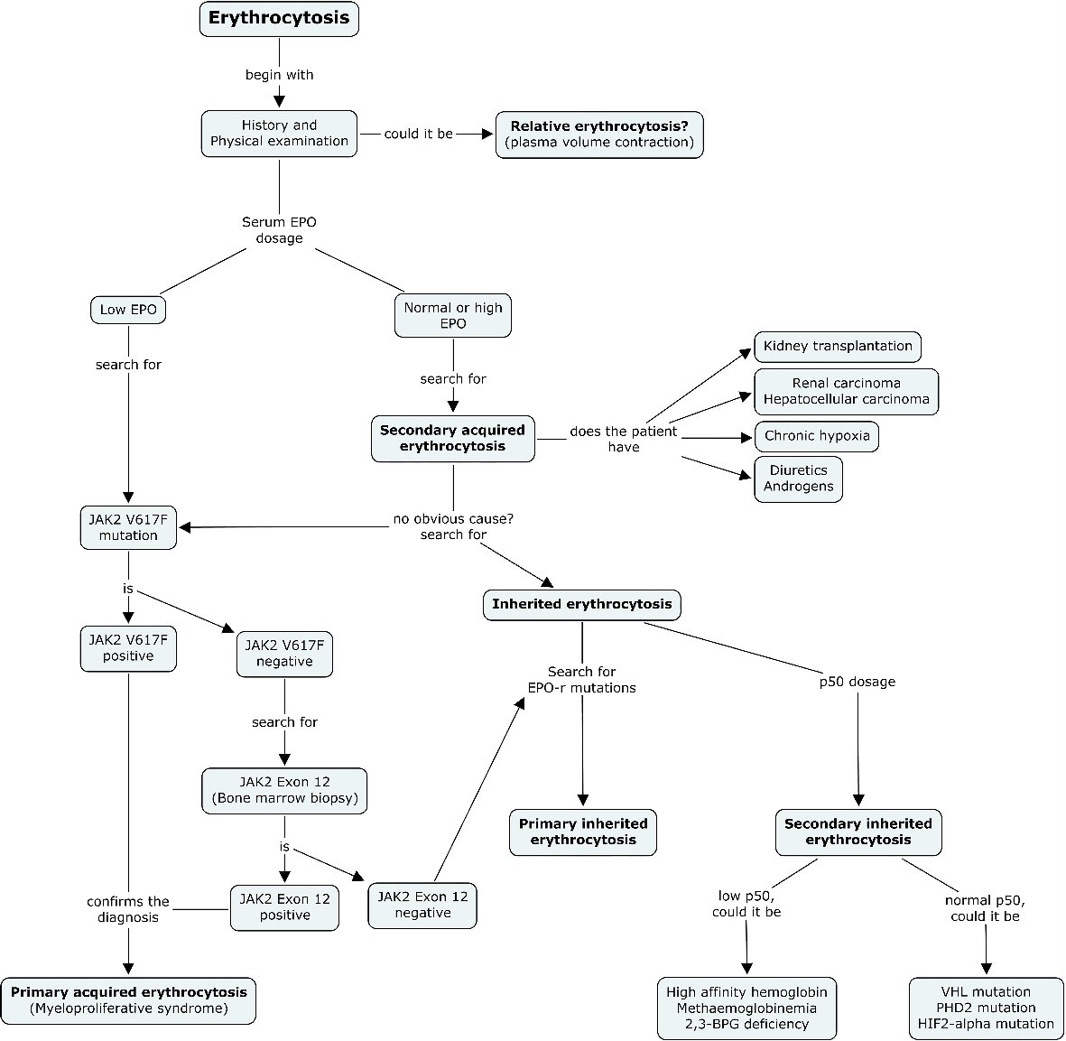
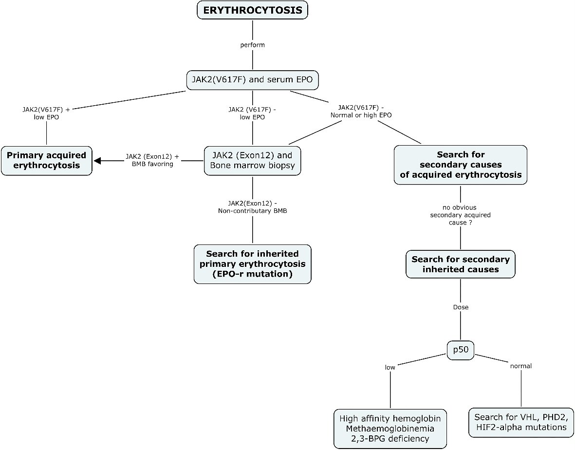
Figure 1. Diagnostic algorithm suggested for erythrocytosis. The current diagnostic approach recommended for the investigation of erythrocytosis systematically includes a search for JAK2 (V617F) mutation and serum EPO as the first step in every patient. A positive JAK2 (V617F) result should orient the diagnosis and management towards a diagnosis of primary acquired erythrocytosis. A normal or high EPO should guide the clinician towards the search for a secondary cause of acquired erythrocytosis, and when no cause is found, the search for a secondary inherited cause. A negative result for JAK2 (V617F) mutation and a low EPO level should be further investigated with a bone marrow biopsy and a search for JAK2 (exon12), and when non-contributory, should prompt a search for inherited primary erythrocytosis. EPO: erythropoietin; BPG: bisphosphoglycerate; HIF-2: hypoxia inducible factor 2; PHD2: prolyl hydroxylase domain protein 2; VHL: von Hippel-Lindau.
Figure 2.
Figure 2. New diagnostic algorithm suggested for erythrocytosis. After exclusion of relative erythrocytosis, serum erythropoietin dosage should be undertaken early in the diagnostic course before concluding of a secondary cause for erythrocytosis in the young adult. In the case of low EPO, JAK2 (V617F) mutation should systematically be searched. A bone marrow biopsy for JAK2 exon 12 search should be undertaken with a negative JAK2 (V617F) mutation. In the case of normal or high EPO, and when no obvious cause for secondary acquired erythrocytosis could be found, a search for inherited erythrocytosis should be undertaken. A search for EPO-r mutations and P50 dosage must then be taken into consideration to complete the diagnostic evaluation.

Tables

Table 1. Main Etiologies of Erythrocytosis ([1, 3, 4, 7-18])
 
Primary
  Acquired
    Myeloproliferative syndrome (polycythemia vera) [1, 3, 4, 7, 8, 9]
  Hereditary
    EPO r-mutations [10, 11]
Secondary
  Hereditary
    Low P50
      High oxygen affinity hemoglobin [12]
      Congenital methemoglobinemia [13, 14]
      2,3-BPG deficiency [15]
    Normal P50
      Mutation of VHL (Chuvash disease) [16]
      Mutation of PHD2 [17]
      Mutation of HIF2-α [18]
  Acquired
    Hypoxia
      Smoking
      Carbon monoxide intoxication
      Living in high altitude
      Lung disease
      Obstructive sleep apnea syndrome
      Intracardiac shunt and cyanotic heart disease
    Renal impairment
      Renal artery stenosis
      Renal cyst
      Post kidney transplant
    Neoplasia
      Renal carcinoma
      Hepatocellular carcinoma
      Cerebellum hemangioblastoma
      Pheochromocytoma
      Uterine leiomyoma
    Endocrinopathy
      Cushing disease
    Drugs
      Diuretics
      Exogenous androgens
      Exogenous EPO


Table 2. Distribution of Cases per Year
 
Year of diagnosis (year)Number of cases (n)
19954
19962
19972
19981
19991
20000
20011
20020
20032
20047
20055
20064
20073
20081
20094
20101
20113
20121
20133
20145
20156


Table 3. Demographic and Clinical Characteristics of Patients
 
Characteristics (n = 56)Value
aIntracardiac shunt, cyanotic heart disease. bChronic obstructive pulmonary disease, sleep apnea syndrome. cPost-kidney transplant. dHigh oxygen affinity hemoglobin. eBody mass index (BMI) ≥ 30kg/m2. fUnderweight (BMI < 18.5kg/m2), normal weight (BMI 18.5 - 24.95kg/m2), overweight (BMI 25.0 - 29.95kg/m2), class I obesity (BMI 30.0 - 34.95kg/m2), class II obesity (BMI 35.0 - 39.95kg/m2), class III obesity (BMI > 40.05kg/m2). *Data of six patients missed.
Age at diagnosis, median value (extremes)27 (16 - 35)
Gender
  Female, n (%)21 (37.5)
  Male, n (%)35 (62.5)
Comorbidities
  Heart diseasea, n (%)4 (7.1)
  Lung diseaseb, n (%)5 (8.9)
  Kidney diseasec, n (%)6 (10.7)
  Hemoglobinopathyd, n (%)2 (3.6)
  Obesitye*, n (%)23 (41.1)
Lifestyle habits
  Tobacco use, n (%)33 (58.9)
  Alcohol, n (%)6 (10.7)
  Recreational drugs, n (%)15 (26.8)
Body mass indexf*, median value (extremes)28.3 (13.5 - 57.0)
  Underweight, n (%)4 (7.1)
  Normal weight, n (%)13 (23.2)
  Overweight, n (%)9 (16.1)
  Obesity - class I, n (%)14 (25.0)
  Obesity - class II, n (%)3 (5.4)
  Obesity - class III, n (%)7 (12.5)
Drugs
  Exogenous androgens, n (%)4 (7.1)
  Anabolic steroids, n (%)0 (0)
  Diuretics, n (%)1 (1.9)
  Exogenous erythropoietin, n (%)0 (0)


Table 4. Erythrocytosis Criteria
 
MenWomen
Hemoglobin, median value
  At diagnosis (g/L)186170
  Highest value during follow-up (g/L)194177
Hematocrit, median value
  At diagnosis (%)53.249
  Highest value during follow-up (%)55.551.6


Table 5. Major Etiologies of Erythrocytosis
 
Etiologies (n = 56)Value
Relative erythrocytosis, n (%)7 (12.5)
Absolute erythrocytosis, n (%)25 (44.6)
  Primary, n (%)0 (0)
  Secondary, n (%)25 (44.6)
No identifiable cause, n (%)24 (42.9)


Table 6. Detailed Etiologies of Secondary Erythrocytosis Cases
 
Value
Hereditary, n (%)
  Low P50, n (%)2 (3.6)
  Normal P50, n (%)0 (0)
Acquired, n (%)
  Hypoxia, n (%)13 (23.2)
  Renal impairment, n (%)6 (10.7)
  Neoplasia, n (%)0 (0)
  Endocrinopathy, n (%)0 (0)
  Drugs, n (%)4 (7.1)


Table 7. Patients Management
 
Tests and analysesNumber of patientsAbnormal values
aAST, ALT, and bilirubin. bVHL, PDH2, HIF-α, EPO-r.
Clinical history and physical examination, n (%)56 (100)-
Oxygen saturation, n (%)51 (91.1)7 (12.5)
Complete blood count, n (%)56 (100)56 (100)
Red blood cell mass, n (%)4 (7.1)3 (5.4)
Liver function testsa, n (%)53 (94.6)18 (32.1)
Chest X-ray, n (%)48 (85.7)9 (16.1)
Abdominal echography, n (%)32 (57.1)12 (21.4)
Search for JAK2 mutation, n (%)10 (17.9)0 (0)
Search for other genetic mutationsb, n (%)0 (0)0 (0)
Serum EPO, n (%)13 (23.2)0 (0)
Bone marrow aspirate and biopsy, n (%)2 (3.6)1 (1.8)
P50, n (%)0 (0)0 (0)
Carboxyhemoglobin, n (%)12 (21.4)0 (0)