Journal of Hematology, ISSN 1927-1212 print, 1927-1220 online, Open Access
Article copyright, the authors; Journal compilation copyright, J Hematol and Elmer Press Inc
Journal website http://www.thejh.org

Case Report

Volume 8, Number 4, December 2019, pages 160-164


Delayed Presentation of Thymoma-Related Aplastic Anemia: An Unusual Presentation of a Rare Complication

Figures

Figure 1.
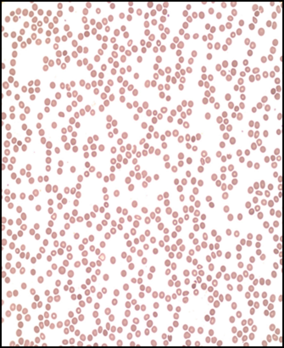
Figure 1. Peripheral blood smear. Markedly reduced red blood cells, platelets, and leukocytes. Mild anisocytosis without significant morphological abnormalities.
Figure 2.
Figure 2. Bone marrow biopsy. The bone marrow shows profound hypocellularity of 5% (expected for age 25-30%) with decreased numbers in all hematopoietic lineages.
Figure 3.
Figure 3. Bone marrow aspirate. There was no evidence of dysplasia. Residual cells including few lymphocytes, plasma cells, and macrophages, some containing hemosiderin, were seen in the bone marrow fat.
Figure 4.
Figure 4. Chest X-ray: 8.0 × 7.5 cm left anterior mediastinal mass.
Figure 5.
Figure 5. CT of the chest: interval enlargement of anterior mediastinal mass measuring 7.8 × 6.9 cm. CT: computed tomography.