Journal of Hematology, ISSN 1927-1212 print, 1927-1220 online, Open Access
Article copyright, the authors; Journal compilation copyright, J Hematol and Elmer Press Inc
Journal website https://www.thejh.org

Original Article

Volume 9, Number 4, December 2020, pages 123-131


Treatment-Related Mortality From Infectious Complications in an Acute Leukemia Clinic

Figures

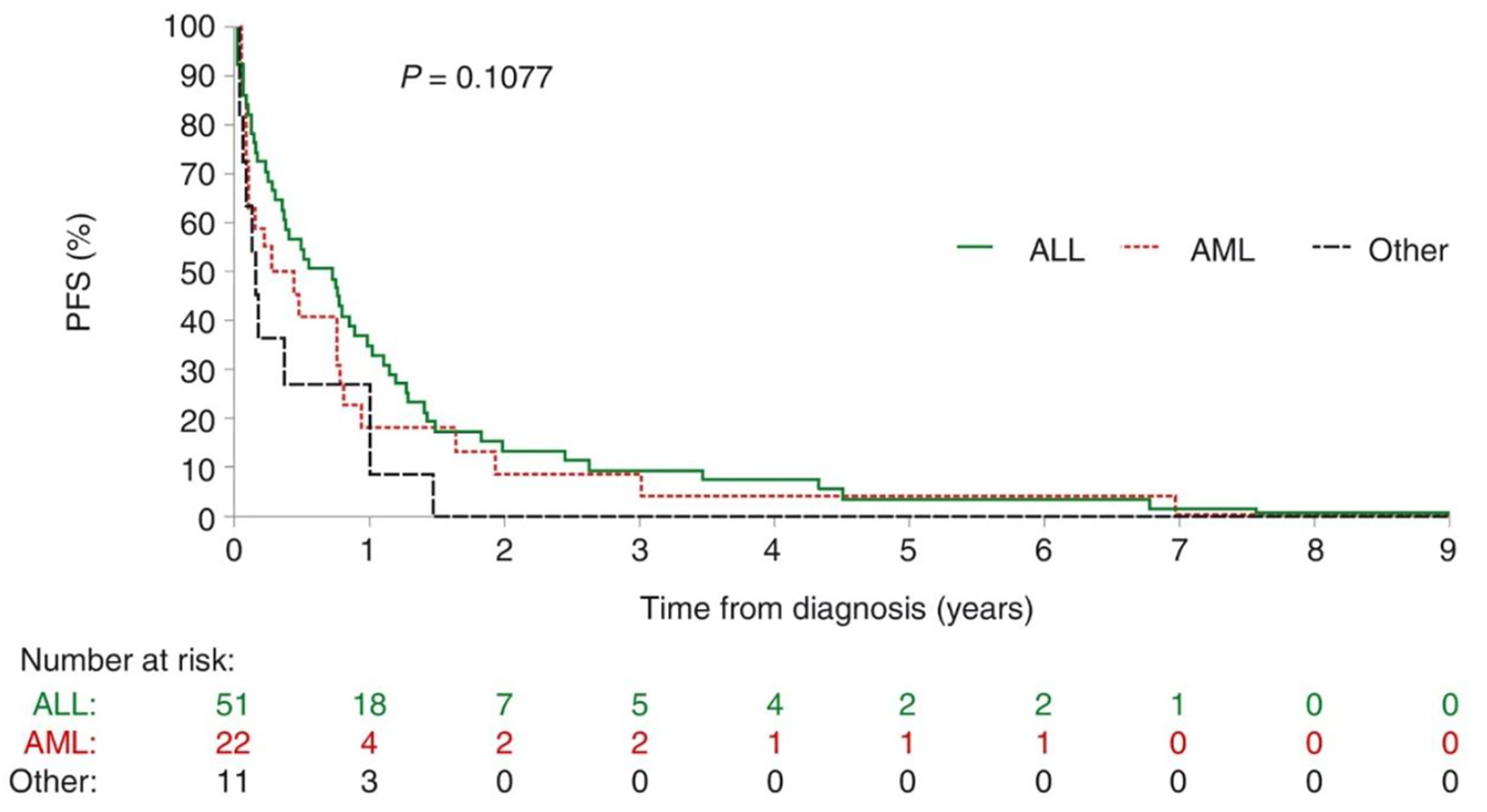
Figure 1.
Figure 1. Progression-free survival in patients with acute leukemia who died of infectious complications. There is no difference between the types of leukemias. ALL: acute lymphoblastic leukemia; AML: acute myeloblastic leukemia; PFS: progression-free survival.
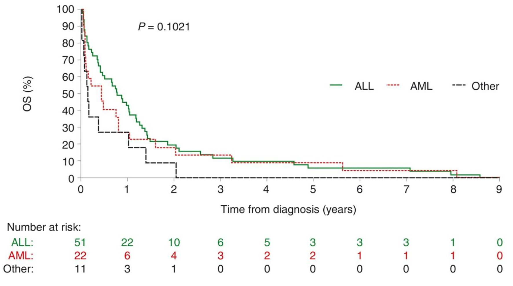
Figure 2.
Figure 2. Overall survival in patients with acute leukemia who died of infectious complications. There is no statistical difference between the two types of leukemias, no difference between relapse or naive leukemia in this Kaplan-Meier curves. A division should be made. ALL: acute lymphoblastic leukemia; AML: acute myeloblastic leukemia; OS: overall survival.

Tables

Table 1. Characteristics of Patients Who Died of Infectious Complications Between January 2012 and December 2015
 
CharacteristicLeukemia typeP value
All types (n = 84)ALL (n = 51)AML (n = 22)Other (n = 11)
aChemotherapy received within 45 days prior to death. AIDA: ATRA + idarubicin; ALL: acute lymphoblastic leukemia; AML: acute myeloblastic leukemia; ATRA: all-trans retinoic acid; BFM: Berlin-Frankfurt-Munster chemotherapy [11]; CALGB: Cancer and Leukemia Group B; Hyper-CVAD: hyperfractionated cyclophosphamide, vincristine, doxorubicin, dexamethasone; IDA-FLAG: idarubicin, fludarabine, high-dose cytarabine, granulocyte colony-stimulating factor; MEC: mitoxantrone, etoposide, intermediate-dose Ara-C; POMP: 6-mercaptopurine, vincristine, methotrexate, prednisone.
Median age, years (range)35.2 (15.8 - 71.3)27.0 (15.8 - 61.2]45.1 (19.0 - 71.3)40.2 (23.2 - 70.6)0.0004
Male/female, n (%)52 (61.9)/32 (42.5)35 (68.6)/16 (31.4)11 (50.0)/11 (50.0)6 (54.5)/5 (45.5)0.2691
Previous disease, n (%)0.0027
  ALL74 (88.1)48 (94.1)20 (90.9)6 (54.5)
  AML1 (1.2)1 (2.0)00
  Blastic plasmacytoid dendritic cell neoplasm1 (1.2)1 (2.0)00
  Chronic myelogenous leukemia2 (2.4)1 (2.0)01 (9.1)
  Diffuse large B-cell lymphoma1 (1.2)001 (9.1)
  Lung cancer1 (1.2)001 (9.1)
  Myelodysplastic syndrome3 (3.6)01 (4.5)2 (18.2)
  Thyroid carcinoma1 (1.2)01 (4.5)0
Relapsed leukemia, n (%)43 (51.2)22 (43.1)12 (54.5)9 (81.8)0.0673
Treatment stage at time of death, n (%)
  Induction27 (32.1)14 (27.5)7 (31.8)6 (54.5)
  Re-induction5 (6.0)3 (5.9)1 (4.5)1 (9.1)
  First relapse25 (29.8)15 (29.4)8 (36.4)2 (18.2)
  Second relapse6 (7.1)4 (7.8)1 (4.5)1 (9.1)
  Consolidation9 (10.7)6 (11.8)2 (9.1)1 (9.1)
  Maintenance2 (2.4)2 (3.9)00
  Palliative care1 (1.2)01 (4.5)0
  Refractory leukemia9 (10.7)7 (13.7)2 (9.1)0
Chemotherapy regimena, n (%)0.2795
  Hyper-CVAD17 (20.2)16 (31.4)01 (9.1)
  7+310 (11.9)1 (2.0)6 (27.3)3 (27.3)
  Palliative POMP10 (11.9)8 (15.7)02 (18.2)
  BFM8 (9.5)8 (15.7)00
  Cytarabine high dose5 (6.0)05 (22.7)0
  Larson regimen [9]5 (6.0)5 (9.8)00
  CALGB regimen [10]1 (1.2)1 (2.0)00
  Transplant4 (4.8)3 (5.9)1 (4.5)0
  AIDA1 (1.2)001 (9.1)
  IDA-FLAG6 (7.1)3 (5.9)3 (13.6)0
  MEC1 (1.2)01 (4.5)0
  Vincristine/dexamethasone3 (3.6)2 (3.9)01 (9.1)
  Steroids1 (1.2)1 (2.0)00
  Blood transfusion6 (7.1)2 (3.9)2 (9.1)2 (18.2)


Table 2. Site/Nature of Infection in Patients Who Died of Infectious Complications
 
Site/nature of infection, n (%)Leukemia type
All types (n = 84)ALL (n = 51)AML (n = 22)Other (n = 11)
The site of infection was documented by site of culture and image studies performed to the patients previously to the death (blood culture, urine culture, feces culture, skin biopsy, sinus swap and catheter culture). ALL: acute lymphoblastic leukemia; AML: acute myeloblastic leukemia.
Catheter-associated infection6 (7.1)4 (7.8)2 (9.1)0
Gastrointestinal tract8 (9.5)4 (7.8)3 (13.6)1 (9.1)
Kidney3 (3.6)2 (3.9)01 (9.1)
Lung56 (66.7)32 (62.7)16 (72.7)8 (72.7)
Skin9 (10.7)8 (15.7)1 (4.5)0
Paranasal sinus2 (2.4)1 (2.0)01 (9.1)


Table 3. Most Frequently Isolated Organisms From Blood, Urine, Feces, or Bronchoscopy Culture in Patients Who Died of an Infectious Complication, Depending of the Different Type of Chemotherapy and the Number of Treatment in Correlation With the Site and the Microorganism of Infection
 
Organism, n (%)Leukemia type
All types (n = 84)ALL (n = 51)AML (n = 22)Other (n = 11)
ALL: acute lymphoblastic leukemia; AML: acute myeloblastic leukemia; ESBL: extended-spectrum beta-lactamases.
None isolated29 (34.5)19 (37.3)7 (31.8)3 (27.3)
Klebsiella oxytoca1 (1.2)1 (2.0)00
Staphylococcus aureus1 (1.2)1 (2.0)00
ESBL-producing Escherichia coli12 (14.3)8 (15.7)2 (9.1)2 (18.2)
Acinetobacter baumannii2 (2.4)01 (4.5)1 (9.1)
Enterococcus faecium7 (8.3)5 (9.8)2 (9.1)0
Pseudomonas aeruginosa5 (6.0)2 (3.9)3 (13.6)0
Stenotrophomonas maltophilia3 (3.6)2 (3.9)1 (4.5)0
Candida albicans4 (4.8)3 (5.9)1 (4.5)0
Escherichia coli2 (2.4)01 (4.5)1 (9.1)
Klebsiella pneumoniae4 (4.8)2 (3.9)1 (4.5)1 (9.1)
Staphylococcus epidermis2 (2.4)002 (18.2)
Candida glabrata2 (2.4)1 (2.0)01 (9.1)
Aeromonas hydrophila1 (1.2)1 (2.0)00
Influenza H1N11 (1.2)01 (4.5)0
Aspergillus flavus1 (1.2)1 (2.0)00
Influenza H3N21 (1.2)01 (4.5)0
Serratia marcescens2 (2.4)2 (3.9)00
Clostridium difficile2 (2.4)1 (2.0)1 (4.5)0
Mucormycosis2 (2.4)2 (3.9)00