| Journal of Hematology, ISSN 1927-1212 print, 1927-1220 online, Open Access |
| Article copyright, the authors; Journal compilation copyright, J Hematol and Elmer Press Inc |
| Journal website http://www.thejh.org |
Letter to the Editor
Volume 1, Number 4-5, October 2012, pages 112-112
Bone Infarct Mimicking Infection in a Young Adult Sickler
Ralph Yachouia, b, Pamela Traisaka
aDivision of Rheumatology, Cooper Medical School of Rowan University, USA
bCorreponding author: Ralph Yachoui, Division of Rheumatology, Cooper Medical School of Rowan University, 900 Centennial Blvd Bldg 2 Ste 201,Voorhees, NJ 08043, USA
Manuscript accepted for publication October 4, 2012
Short title: Bone Infarct Mimicking Infection
doi: https://doi.org/10.4021/jh41w
| To the Editor | ▴Top |
A 23-year-old known sickle cell patient presented with left knee swelling and pain of 10 days duration. He was treated several times in the past for painful crises. Currently, at rest the knee pain was 8/10 and upon palpation or movement the pain was 10/10. Moving the knee at all aggravated his symptoms. His temperature was 101. The laboratory investigations demonstrated high white blood cell count at 19,000/µL (normal 4,000 – 11,000/µL) and a hemoglobin level of 8.5 g/dL. Initial plain x-ray was normal. An arthrocentesis yielded 30 cc of yellow dark fluid and synovial fluid analysis revealed 3,000 WBC (80% PMN) with no crystals. Final culture was unrevealing.
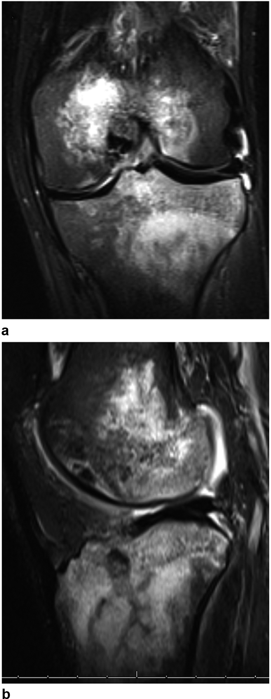
A magnetic resonance imaging (MRI) showed increased T2 signal in the distal femur and proximal tibia (Fig. 1). The fatty yellow marrow in the distal femur and proximal tibia was replaced with hematopoietic red marrow. These findings indicated bone edema and acute bone infarction.
 Click for large image | Figure 1. Magnetic resonance imaging showed increased T2 signal in the distal femur and proximal tibia (a, b). |
Patients with sickle cell anemia are prone to both infarctive and infective crises. Clinical differentiation between both can be difficult. Initial radiographs are usually normal with an acute infaction [1]. As the condition becomes more chronic sclerosis develops [1]. MRI has an important role in demonstrating the area of infarct in the earlier stages [2]. Bone infarcts of the knees are not as common as in the hips and shoulders [2]. Thus, it is important to consider infarctions in such atypical sites as a differential diagnosis of painful knee pain in sicklers.
Disclosure
None.
| References | ▴Top |
- Ganguly A, Boswell W, Aniq H. Musculosqueletal manifestations of sickle cell anaemia: a pictorial review. Anemia. 2011;2011:794283.
doi pubmed - Ejindu VC, Hine AL, Mashayekhi M, Shorvon PJ, Misra RR. Musculoskeletal manifestations of sickle cell disease. Radiographics. 2007;27(4):1005-1021.
doi pubmed
This is an open-access article distributed under the terms of the Creative Commons Attribution License, which permits unrestricted use, distribution, and reproduction in any medium, provided the original work is properly cited.
Journal of Hematology is published by Elmer Press Inc.


