| Journal of Hematology, ISSN 1927-1212 print, 1927-1220 online, Open Access |
| Article copyright, the authors; Journal compilation copyright, J Hematol and Elmer Press Inc |
| Journal website http://www.thejh.org |
Case Report
Volume 1, Number 4-5, October 2012, pages 93-94
Acute Cutaneous Flare and Hemophagocytosis: An Unusual Presentation of Adult Anaplastic Large Cell Lymphoma
Ralph Yachouia, b, Patrick M Cronina
aDivision of Rheumatology, Cooper Medical School of Rowan University, USA
bCorresponding author: Ralph Yachoui, Division of Rheumatology, Cooper Medical School of Rowan University, 900 Centennial Blvd Bldg 2 Ste 201,Voorhees, NJ 08043, USA
Manuscript accepted for publication July 20, 2012
Short title: Acute Cutaneous Flare and Hemophagocytosis
doi: https://doi.org/10.4021/jh35e
| Abstract | ▴Top |
Hemophagocytic lymphohistiocytosis (HLH), also known as hemophagocytic syndrome, is a disorder characterized by the pathologic activation and proliferation of histiocytes, mainly in the bone marrow, liver, spleen and lymph nodes. Although HLH is frequently associated with lymphomas, it is a rare presentation of anaplastic large cell lymphoma (ALCL). We report a case of anaplastic large cell lymphoma (ALCL) in an adult manifested as generalized cutaneous nodules and fulminant hemophagocytosis.
Keywords: Hemophagocytic lymphohistiocytosis; Anaplastic large cell lymphoma
| Introduction | ▴Top |
Hemophagocytic lymphohistiocytosis (HLH) is an aggressive and potentially life-threatening disease most often affecting infants from birth to 18 months of age, but cases in older children and adults have been reported [1]. It is due to cytokine dysfunction causing immune dysregulation. HLH can be inherited or acquired; the latter being most commonly associated with infections, autoimmune disorders, hematological malignancies or immunocompromised states [2].
| Case Report | ▴Top |
A 54-year-old African American man was with a history of chronic ulcerating skin lesions presented with fever, weight loss, and worsening cutaneous ulcers. Two years prior to his presentation, he developed multiple areas of hyperpigmented skin nodules and extensive exophytic plaques. Multiple skin biopsies had shown activated lymphocytes without atypia.
Physical examination showed diffuse hyperkeratotic scaly papules with central ulceration and yellow drainage. Multiple subcentimeter lymph nodes were palpable in the cervical, axillary and inguinal areas. Hemoglobin was 7.8 g/dL, leukocytes 2.0 × 10/L and platelets count 47 × 10/L. Serum ferritin level was 27,591 ng/mL (reference range (15 - 400 ng/mL)). Plasma fibrinogen level was 150 mg/dL (reference range (210 - 634 mg/dL)). A computed tomography scan revealed a homogeneous enlarged liver measuring 21 cm, and enlarged inguinal lymph nodes, the largest measuring 3.6 × 1.2 × 2 cm.
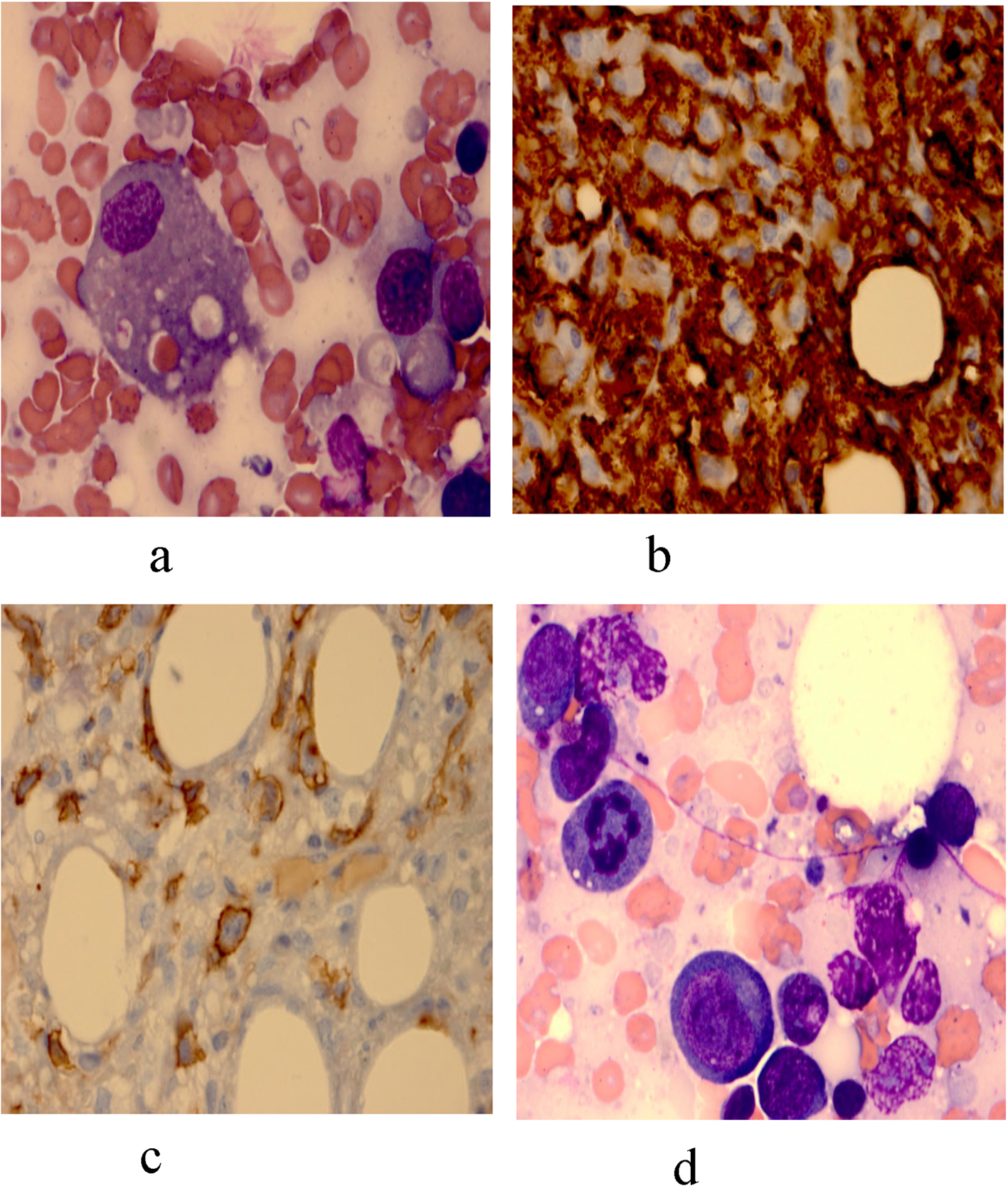
The patient was empirically treated with antibiotics and high dose steroids daily. A bone marrow biopsy demonstrated histiocytic hyperplasia with hemophagocytosis (Fig. 1a), and a diffuse infiltration of atypical lymphoid cells that were CD3-, CD4+, CD30+, ALK-, CD20- and CD163+ (Fig. 1b-d) favoring a diagnosis of anaplastic large cell lymphoma (ALCL). The patient status worsened despite empiric steroid therapy, and he expired of multiorgan failure four weeks after hospitalization.
 Click for large image | Figure 1. (a): Bone marrow aspirate revealing hemophagocytosis; (b): Histiocytic hyperplasia highlighted by CD163 immunostain; (c): The lymphoid cells marked with CD30 immunostain; (d): Anaplastic lymphoma cells, one in mitosis. |
| Discussion | ▴Top |
This is an unusual case of anaplastic large cell lymphoma (ALCL) that presented as generalized cutaneous nodules and fulminant hemophagocytosis. The fever along with pancytopenia, elevated ferritin, hypofibrinogenemia, and hemophagocytosis in the bone marrow confirmed the diagnosis of HLH in our patient. This was the initial presentation of the underlying lymphoproliferative disease. HLH secondary to malignant lymphoma usually manifest with a fulminant clinical course masking the underlying malignancy. Consequently, the possibility of a masked malignant disease should always be considered and ruled out by means of biopsies of involved sites and bone marrow.
HLH secondary to ALCL is extremely uncommon in adults, and only few cases of pediatric HLH secondary to ALCL have been reported [3]. As for the cutaneous aspect of this disease, a single case of diffuse hyperpigmented eruption as a manifestation of systemic ALCL has been described in the literature [4]. Unusual cutaneous lesions in presence of features of bone marrow failure should alert the internist about the possibility of an underlying malignancy.
Disclosure Statement
All authors have no disclosures.
| References | ▴Top |
- Creput C, Galicier L, Buyse S, Azoulay E. Understanding organ dysfunction in hemophagocytic lymphohistiocytosis. Intensive Care Med. 2008;34(7):1177-1187.
pubmed doi - Florena AM, Iannitto E, Quintini G, Franco V. Bone marrow biopsy in hemophagocytic syndrome. Virchows Arch. 2002;441(4):335-344.
pubmed doi - Deborah W, Sevilla MD, John K, Choi MD. Unusual manifestation of pediatric anaplastic large cell lymphoma: Report of two cases. Am J Clin Pathol. 2007; 127: 458-64.
- Masuno Y, Matsumura Y, Katoh M, Arakawa A, Kore-Eda S, Ishikawa T, Miyachi Y. Eosinophilic, polymorphic, and pruritic eruption associated with radiotherapy (EPPER) mimicking bullous pemphigoid in a patient with anaplastic large cell lymphoma. Eur J Dermatol. 2011;21(3):421-422.
pubmed
This is an open-access article distributed under the terms of the Creative Commons Attribution License, which permits unrestricted use, distribution, and reproduction in any medium, provided the original work is properly cited.
Journal of Hematology is published by Elmer Press Inc.


