| Journal of Hematology, ISSN 1927-1212 print, 1927-1220 online, Open Access |
| Article copyright, the authors; Journal compilation copyright, J Hematol and Elmer Press Inc |
| Journal website http://www.thejh.org |
Letter to the Editor
Volume 3, Number 1, March 2014, pages 25-26
Unusual Origin of Anemia With Gluteus Red Spots
Alexander P Rozina, d, Euvgeni Vlodavskyb, Daniel Levinc
aB. Shine Department of Rheumatology, Rambam Health Care Campus and Technion, Haifa, Israel
bDepartment of Pathology, Rambam Health Care Campus and Technion, Haifa, Israel
cDepartment of Orthopedic Surgery, Rambam Health Care Campus and Technion, Haifa, Israel
dCorresponding author: Alexander Rozin, B. Shine Department of Rheumatology, Rambam Health Care Campus and Technion, PO Box 9602, Haifa 31096, Israel
Manuscript accepted for publication December 4, 2013
Short title: Unusual Origin of Anemia With Gluteus Red Spots
doi: https://doi.org/10.14740/jh120e
| To the Editor | ▴Top |
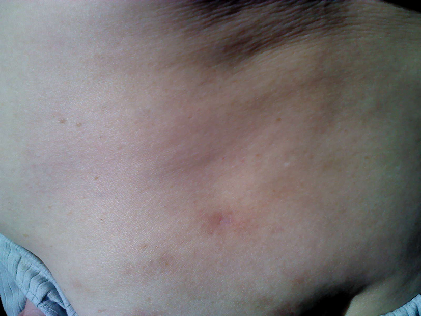
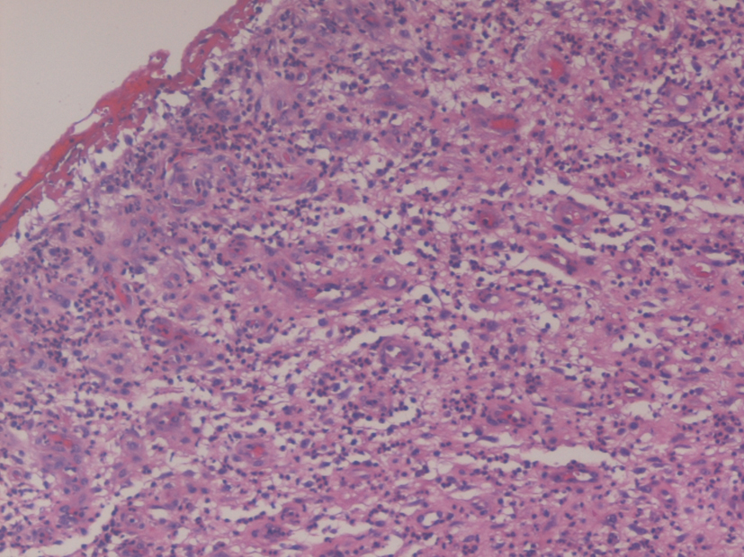
A 62-year-old woman presented with a new history of dizziness, fatigue, scant red gluteal spots and microcytic anemia of 9.9 g/dL. One year and 8 months ago, she underwent total hip replacement with successful rehabilitation. On admission, pallor of skin and red spots of gluteus region on the operation side were seen (Fig. 1). Physical examination of internal organs was unremarkable. Both hip joint motions were within normal limits. Gastro- and colonoscopy did not clarify any origin of anemia. Total body CT showed synovial mass around a hip prosthesis. MRI showed metal-on-metal hip joint transplant with moderate account of synovial fluid and extensive synovial mass distributed around the joint to trochanter pushing back gluteus maximus. Reactive lymphadenopathy was seen along to iliac and groin areas. Final needle biopsy of the periarticular mass was negative for acid fast, non-acid fast bacteria and malignancy and revealed extensive inflammation. All metal implants, and metal-on-metal in particular, corrode and cause a release of metal ions. Chromium, cobalt (N < 0.25 µg/L, patient 15 µg/L) and molybdenum were implicated in development of aseptic lymphocytic vasculitis associated lesions (ALVAL) [1]. The last was proposed as a hypothesis and as a reason of anemia and rash without prosthetic failure [2]. Next step was operative revision. Synovial mass was removed and showed typical lymphocytic vasculitis inflammatory tissue (Fig. 2) [3]. The metal-on-metal prosthesis was replaced to ceramic-to-ceramic device. Rapid recovery, disappearance of vasculitis and restoration of hemoglobin (12.3 g/dL) followed. Anemia was due to chronic inflammation and the metal mediated toxiciy.
 Click for large image | Figure 1. Scarce gluteus red spots on the operation side are seen indicating distribution of synovial inflammation. |
 Click for large image | Figure 2. Fibrous and granulation tissue with marked inflammation and fibrin deposition on the surface. Lymphocytic vasculitis of small vessels is seen. Hematoxylin and eosin, × 200. |
Conflict of Interests
None.
| References | ▴Top |
- Witzleb WC, Ziegler J, Krummenauer F, Neumeister V, Guenther KP. Exposure to chromium, cobalt and molybdenum from metal-on-metal total hip replacement and hip resurfacing arthroplasty. Acta Orthop. 2006;77(5):697-705.
doi pubmed - Reito A, Puolakka T, Pajamaki J. Birmingham hip resurfacing: five to eight year results. Int Orthop. 2011;35(8):1119-1124.
doi pubmed - Campbell P, Ebramzadeh E, Nelson S, Takamura K, De Smet K, Amstutz HC. Histological features of pseudotumor-like tissues from metal-on-metal hips. Clin Orthop Relat Res. 2010;468(9):2321-2327.
doi pubmed
This is an open-access article distributed under the terms of the Creative Commons Attribution License, which permits unrestricted use, distribution, and reproduction in any medium, provided the original work is properly cited.
Journal of Hematology is published by Elmer Press Inc.


